
School of Innovation & Technology
/ MSc Medical Visualisation & Human Anatomy
/ Wei Lin
/ Volumetric Visualisation
Volumetric Visualisation

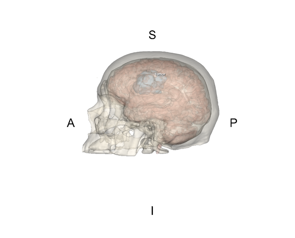
Brain Tumor Identification
Pelvis_Fracture_Visualization
The following works explore how medical scan data can be transformed into clear and informative 3D visualisations. Using CT, MRI and PET datasets in 3D Slicer and MITK, the following were created: pre and post-operative pelvic fracture, internal tooth anatomy, a brain tumour and its surrounding structures, and lung carcinoma. Direct and indirect volume rendering, image registration, filtering, segmentation, surface modelling and colour coding were used to reveal anatomical structure, density, and spatial relationships between the structures.
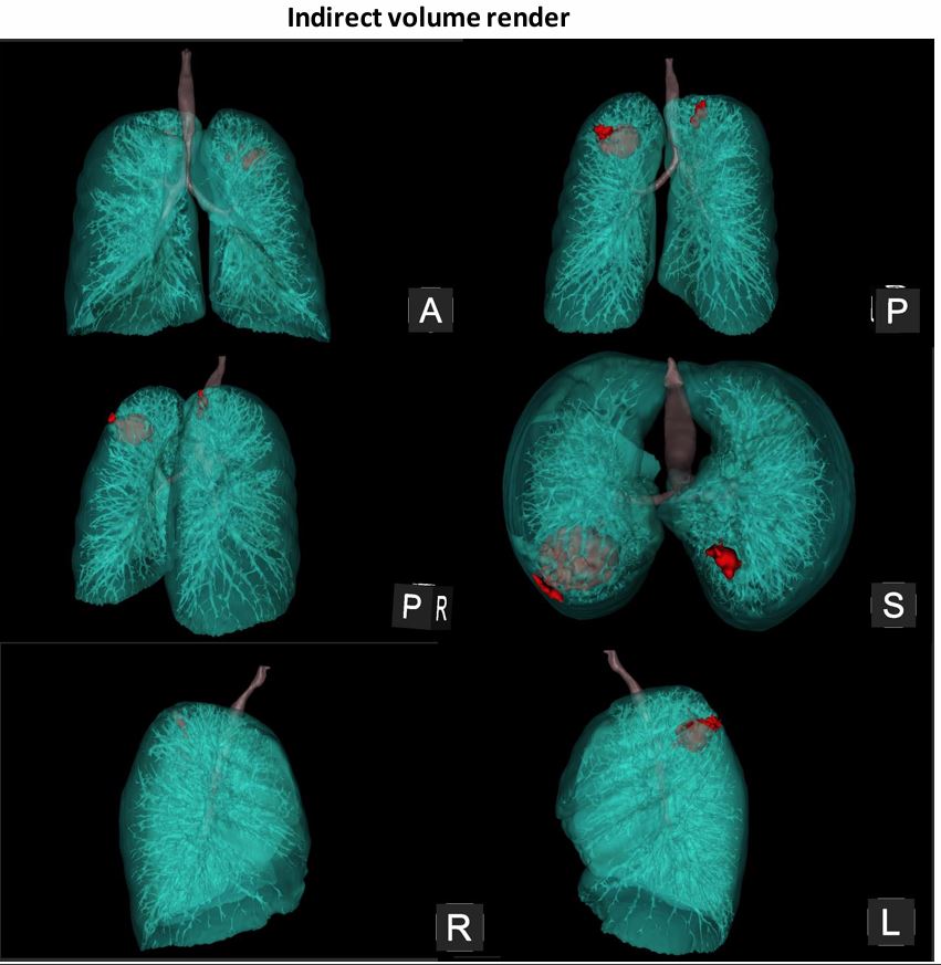
Lungs and tumor were segmented from a CT scan
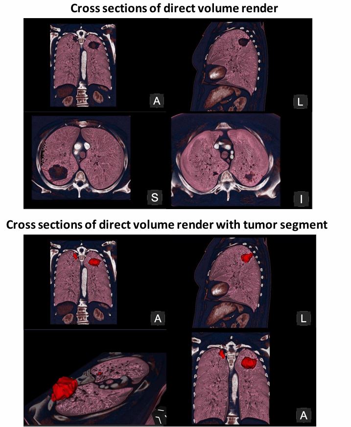
Lungs and tumor were segmented from a CT scan through direct and indirect volume rendering
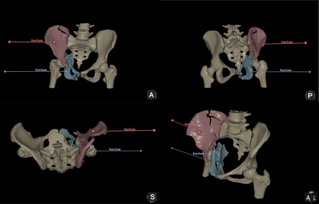
Pre-operative visualisation of a Pelvic Fracture
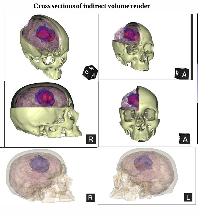
Direct and Indirect Volume Renders
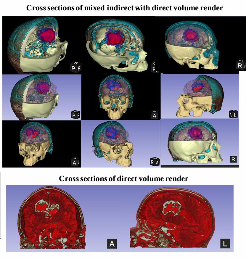
Direct and Indirect Volume Renders
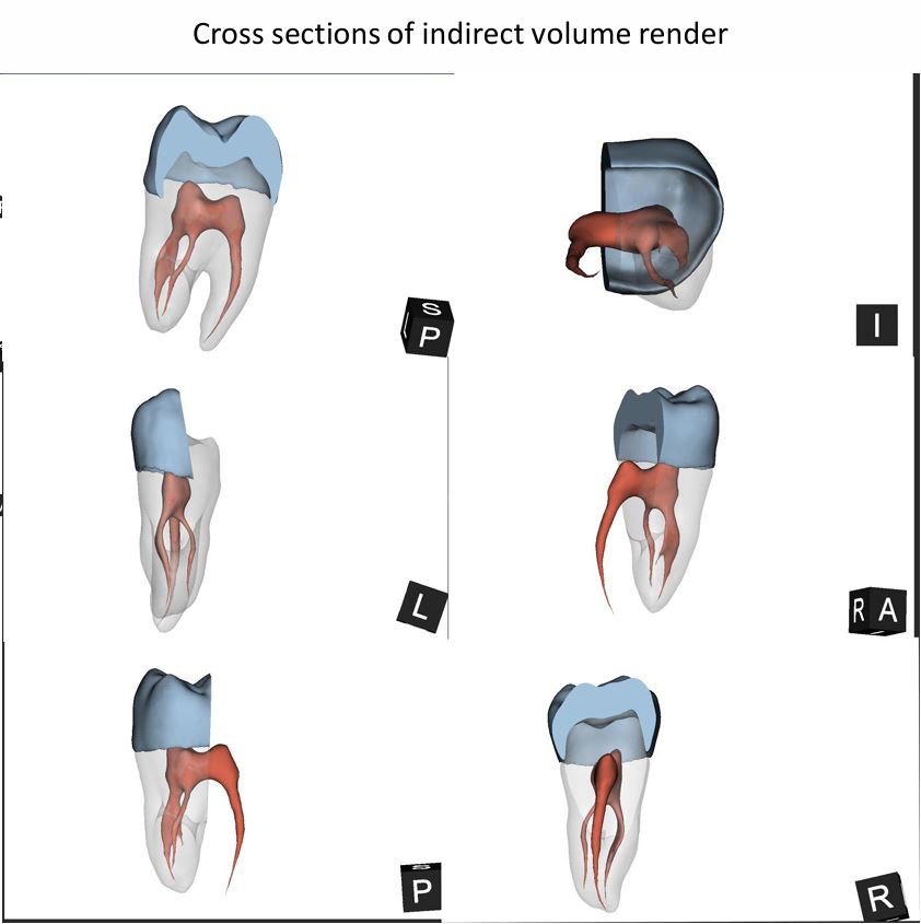
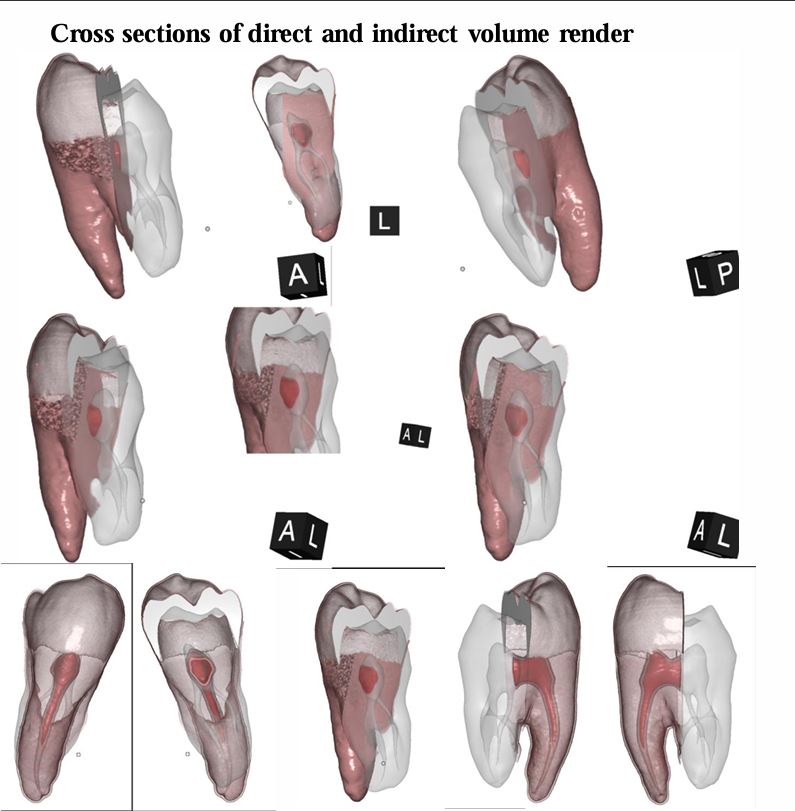
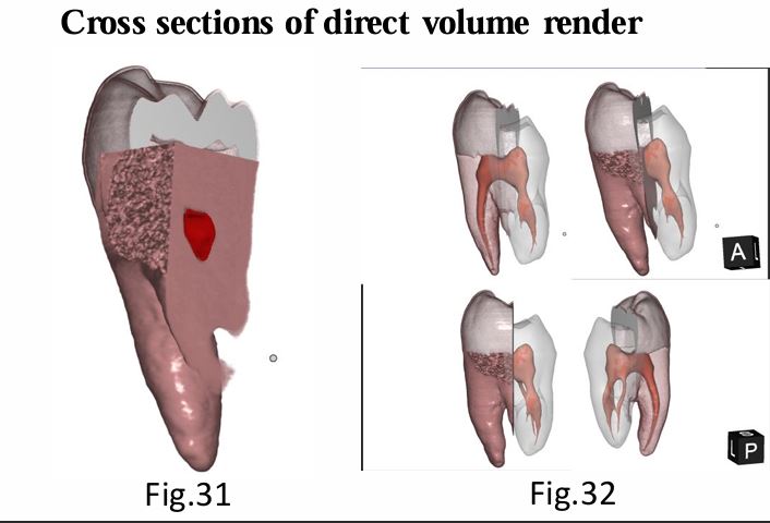
CT scan Segmented Tooth
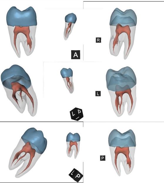
CT scan Segmented Tooth
Segmented and registered the given CT and PET data and rendered them into 3D models to showcase the data for patients
What did I do:
Result: Four rendered outputs of four different anatomies and their datasets.
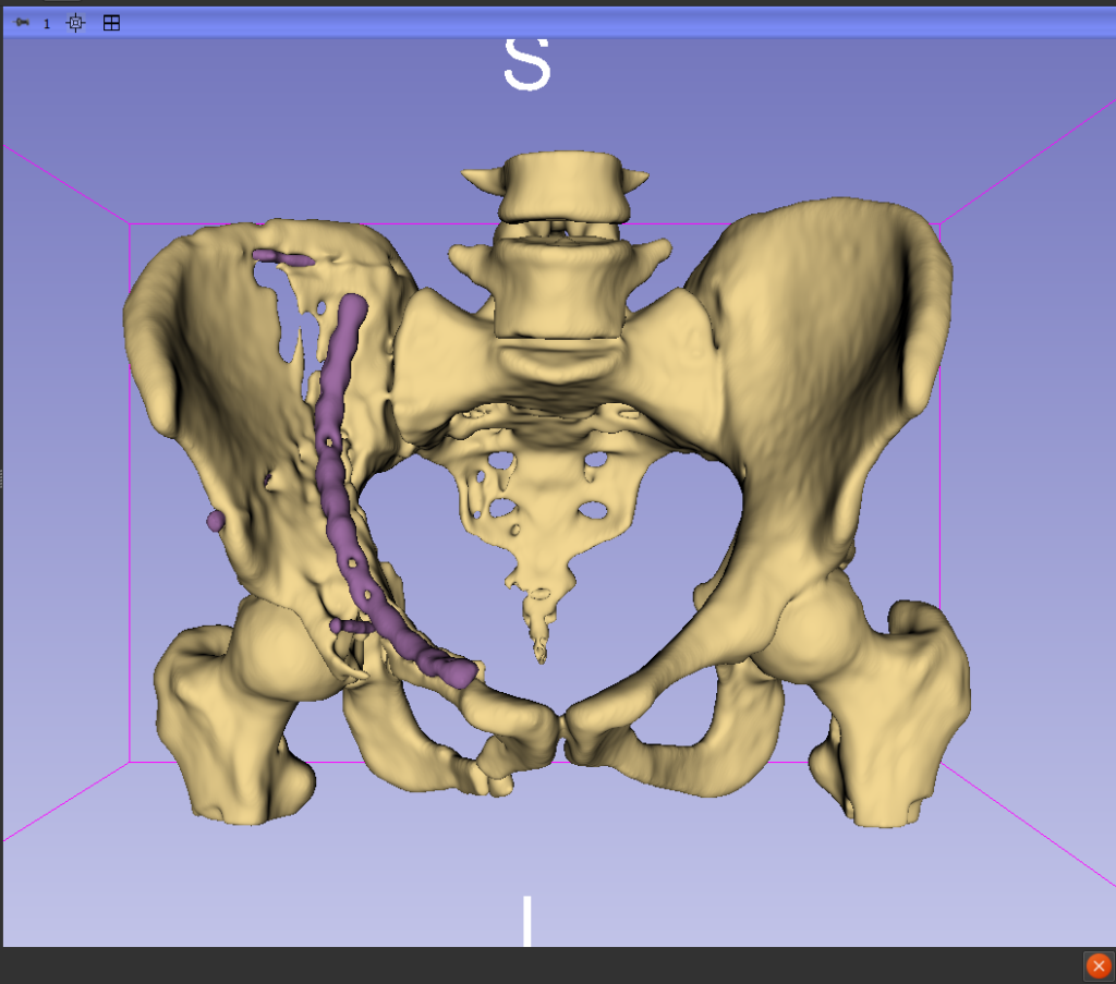
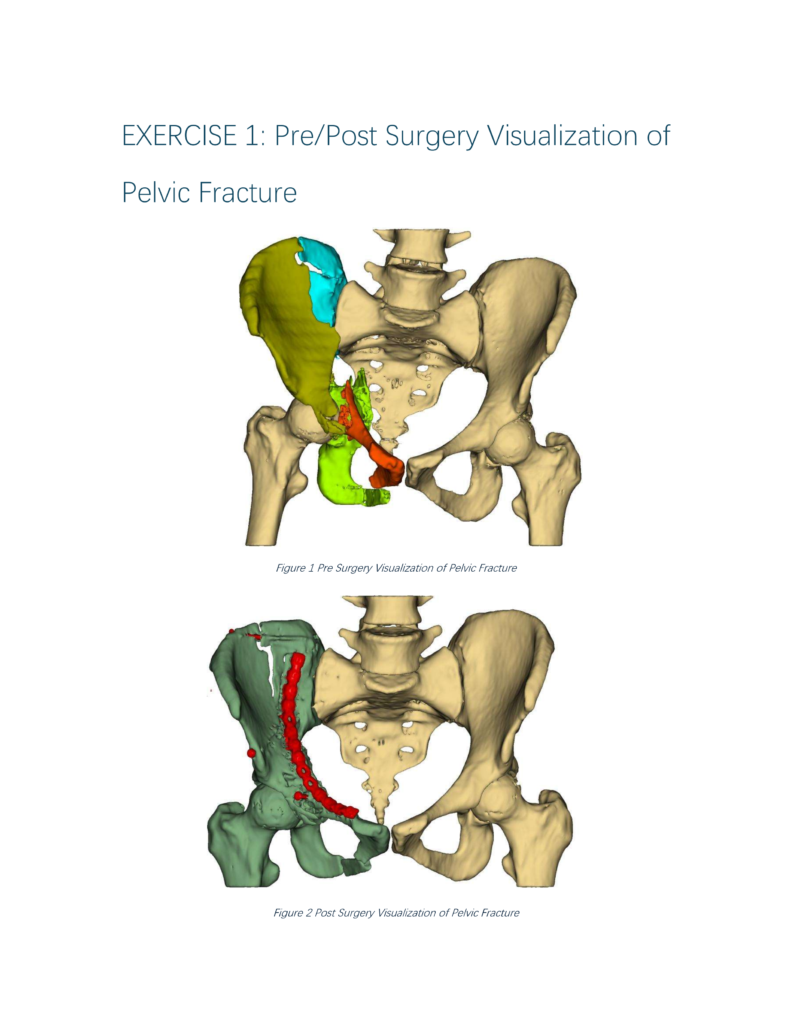
Post op Pelvic bone segmented showing the Implant
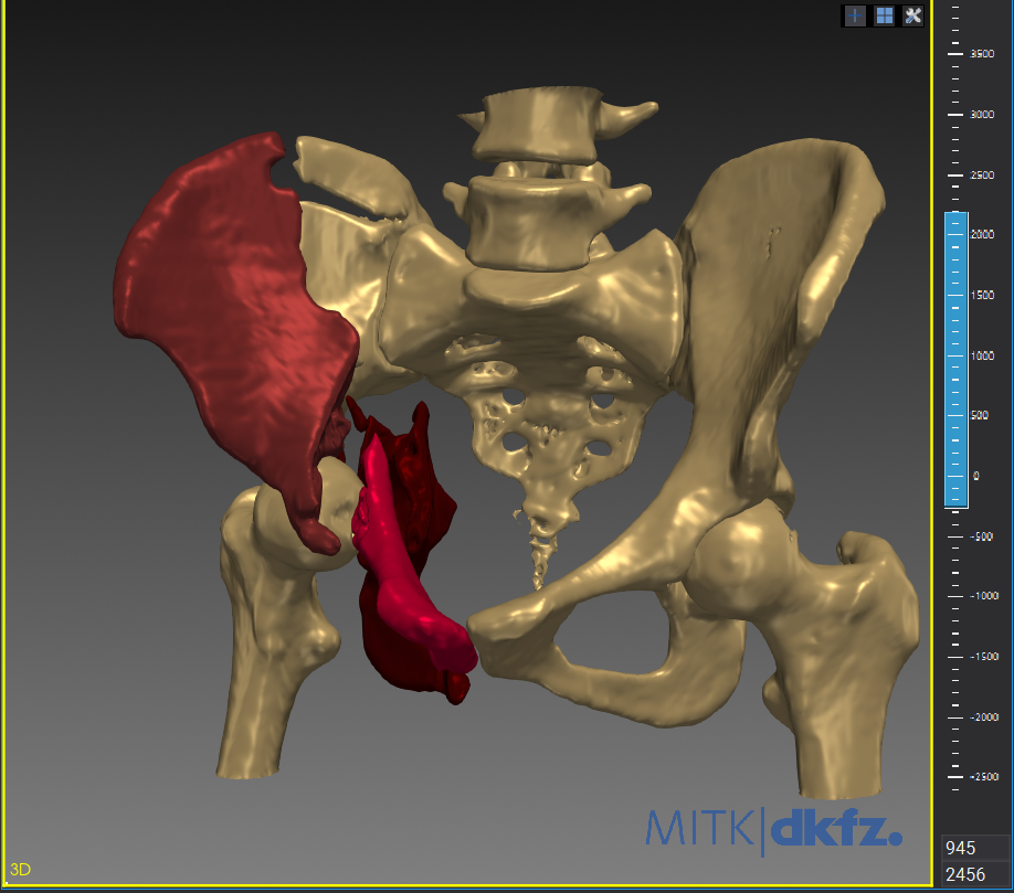
Pre Op Pelvic bone showing the fractures segments
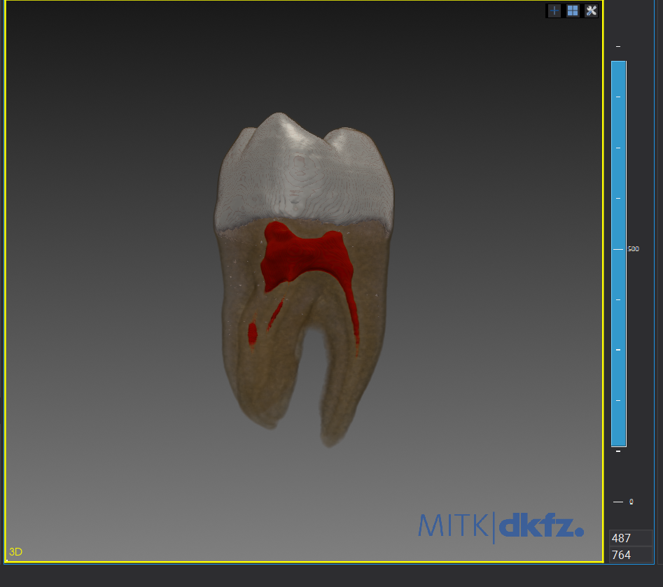
Direct Volume Rendering of Teeth
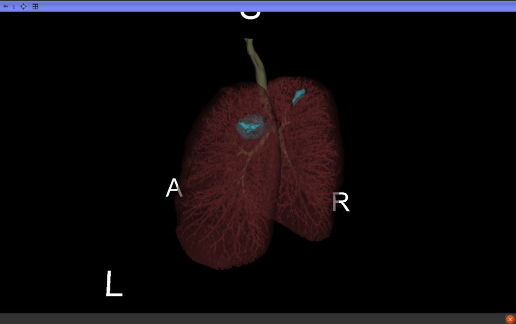
Lungs showing tumor growth on both the upper lobes
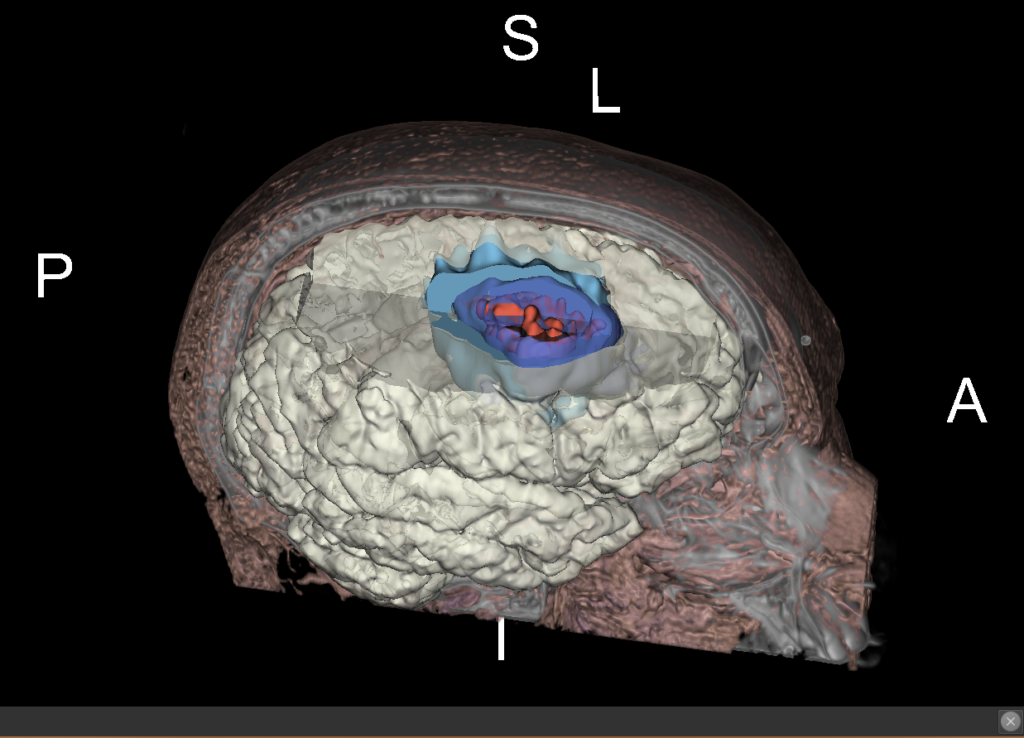
Brain Tumor showing the growth, vasculature, metastasis and borders
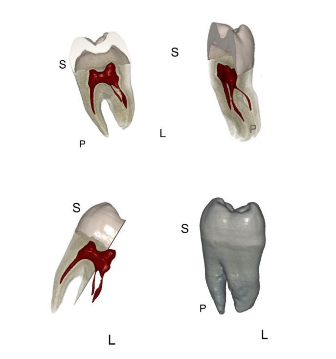
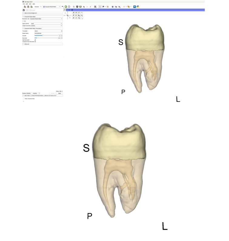
The following images were all from the volumetric visualisation course during semester one. These were done using 3DSlicer and MITK.
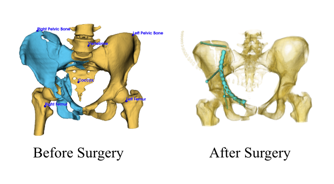
A fractured pelvis is seen on the left before surgery. The pelvis on the right shows the metal that was added to the pelvic fracture after surgery.
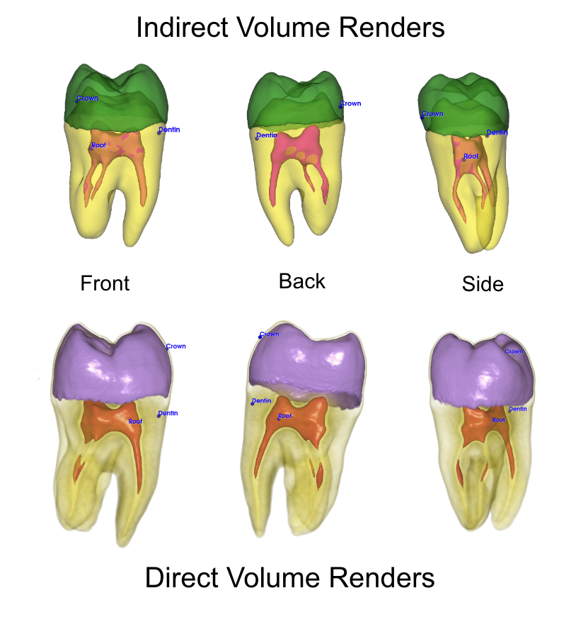
Shown are three different views of indirect and direct volume renders of a tooth.
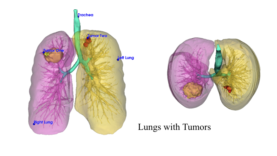
Shown are a pair of lungs with a tumor located in each.
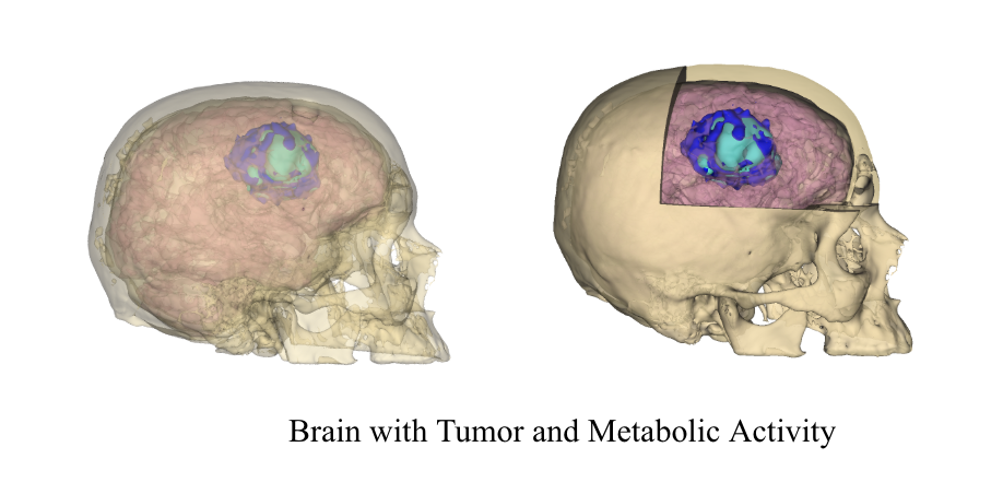
Shown is a skull with a brain containing a tumor and its metabolic activity.