Volumetric Visualisation

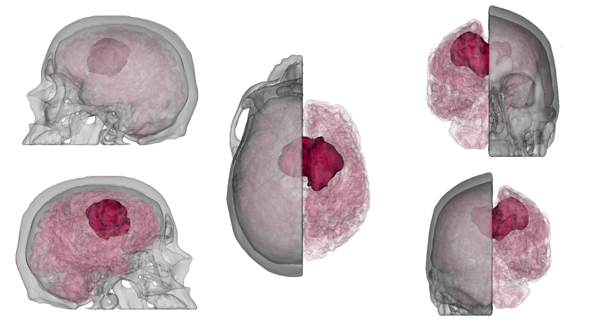
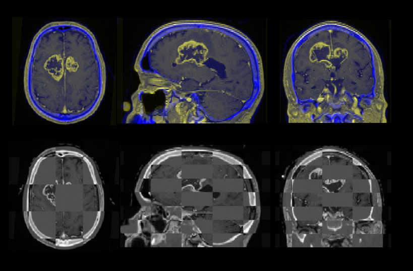
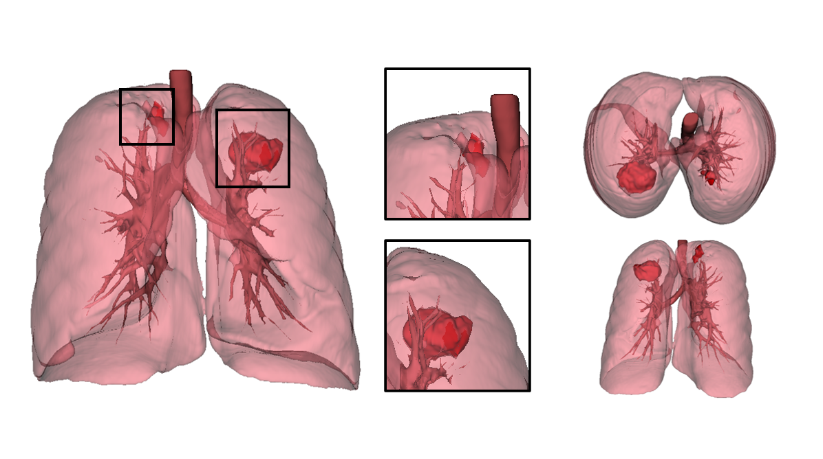
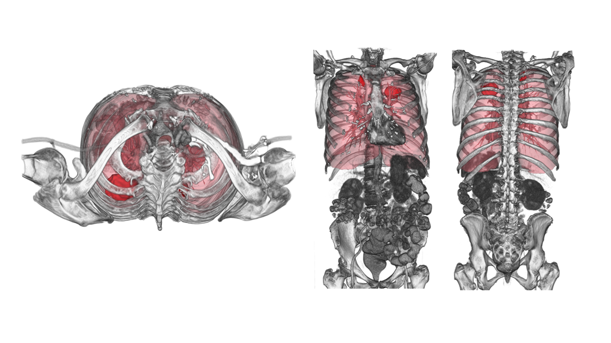
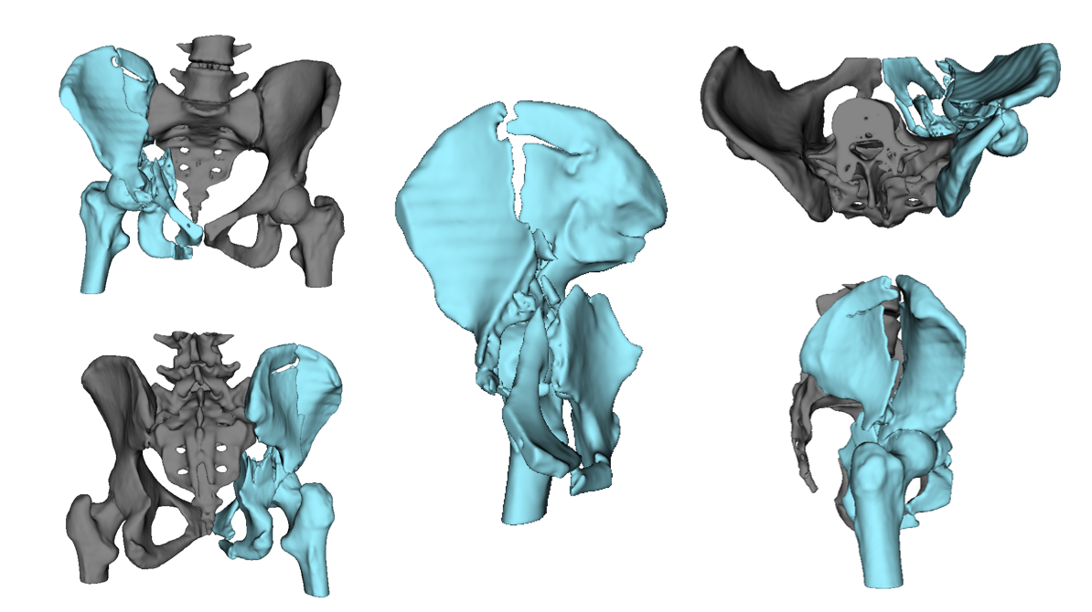
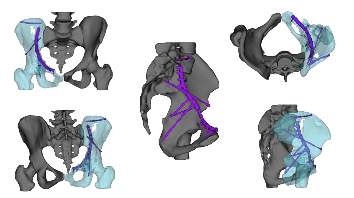
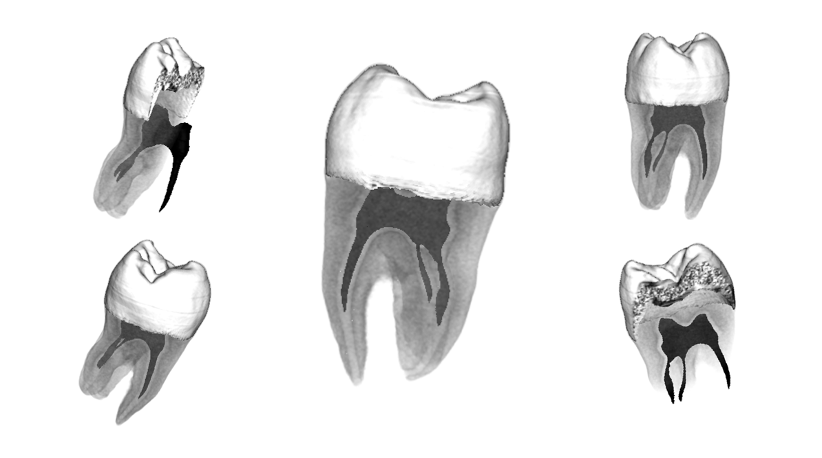
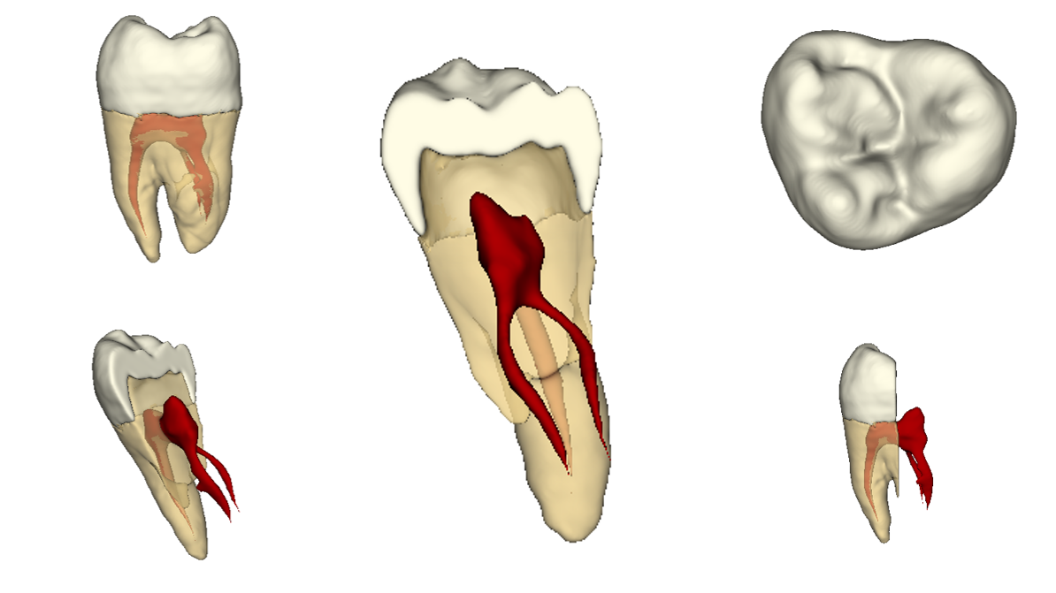
In this module we used 3DSlicer and MITK to visualise anatomy from MRI and CT imaging datasets. The final project required techniques including dataset registration, direct and indirect volume rendering, to produce 3D-visualisations for different audiences.

Brain Tumour Visualisation

The brain and skull were visualised separately and combined to visualise the location of the tumour. This was aimed at a technical audience, to visualise more complex anatomy and used PET, CT and MRI datasets.

Dataset Registration

This shows the dataset registration process using PET, CT and MRI datasets, for the visualisation of the tumour.

Lung Tumours– Identification

The lungs, trachae and bronchial tree were segmented and made transparent to visualise the location of each tumour.

Lung Tumours– Anatomical Relations

To put the lung visualisation in context, the surrounding anatomy was visualised using direct volume rendering.

Pelvic Fracture Visualisation

Pre-operative visualisation of pelvic fractures for a patient.

Post-Op Visualisation of the Pelvis

This visualisation was for a patient post-surgery, to visualise the surgical changes within their pelvis anatomy.

Tooth– Direct Volume Rendering

Showing greater anatomical accuracy of the tooth due to the varying iso-densities between structures.

Tooth– Indirect Volume Rendering

Each structure was segmented individually, providing a cleaner appearance.