MSc Medical Visualisation & Human Anatomy School of Innovation & Technology
Thitiorn Ussavarangsi

My name is Thitiorn Ussavarangsi (Orn). After earning my bachelor’s degree in veterinary medicine and working as a veterinarian for four years in Thailand, my passion has always been about arts.
Throughout my working journey, I learned that effective communication and teamwork are fundamental to meaningful work. This experience, combined with my expertise for animal health sciences, led me to pursue a Master’s degree in Medical Visualisation and Human Anatomy programme.
This course has opened new territories for me. I learned beyond 2D visualisations and embraced cutting-edge technologies such as Augmented Reality (AR), and Virtual Reality (VR). Along this journey, I found joy in learning new skills like 3D modeling, interactive application development, and 3D reconstruction of medical datasets.
My work aims to make complex medical and biological concepts more understandable and engaging. Please enjoy!
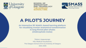
A Pilot’s Journey
Part of animal specimens; Ovaries
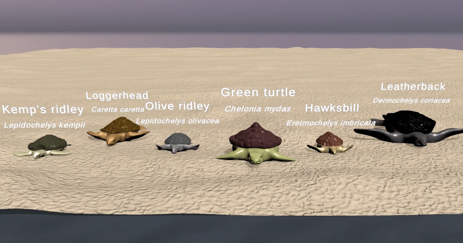
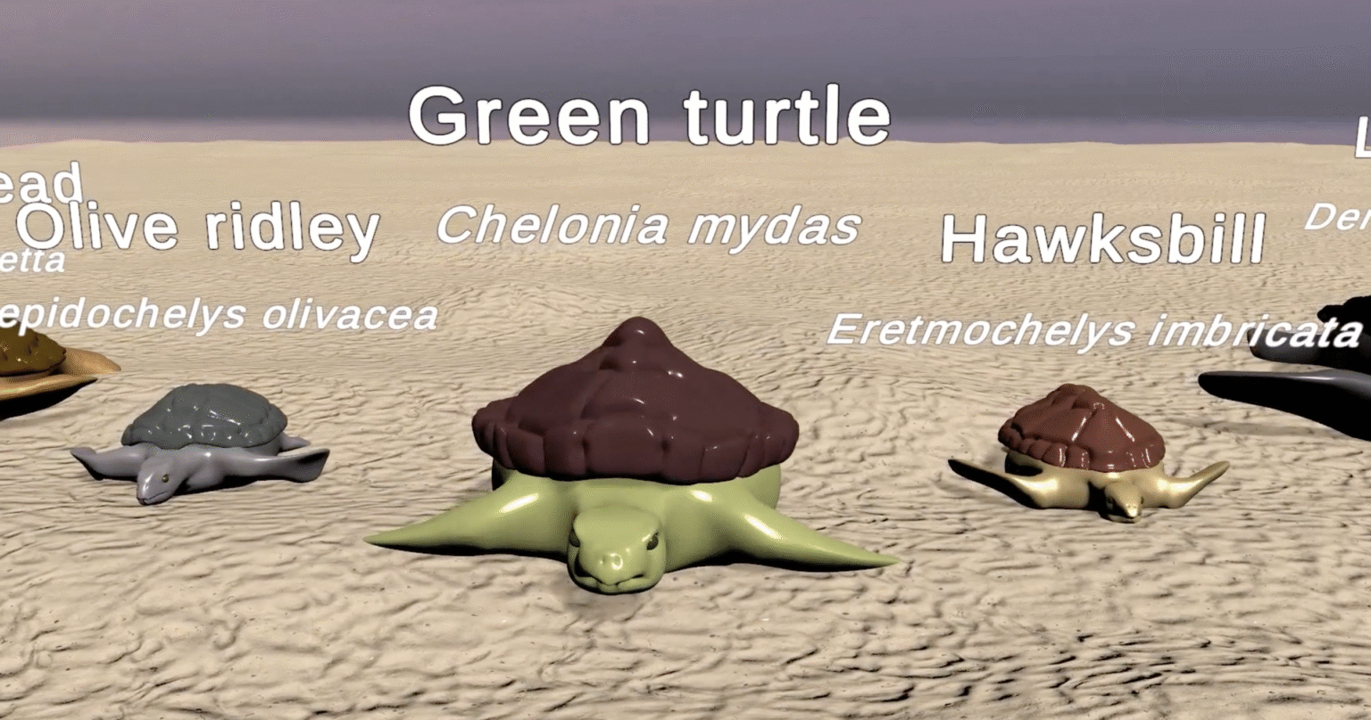
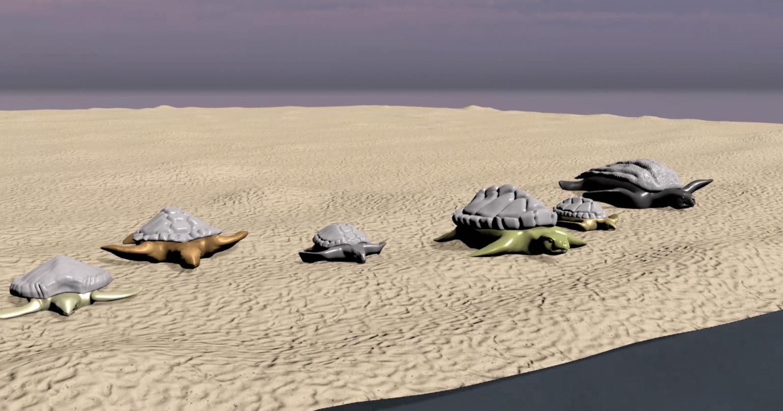
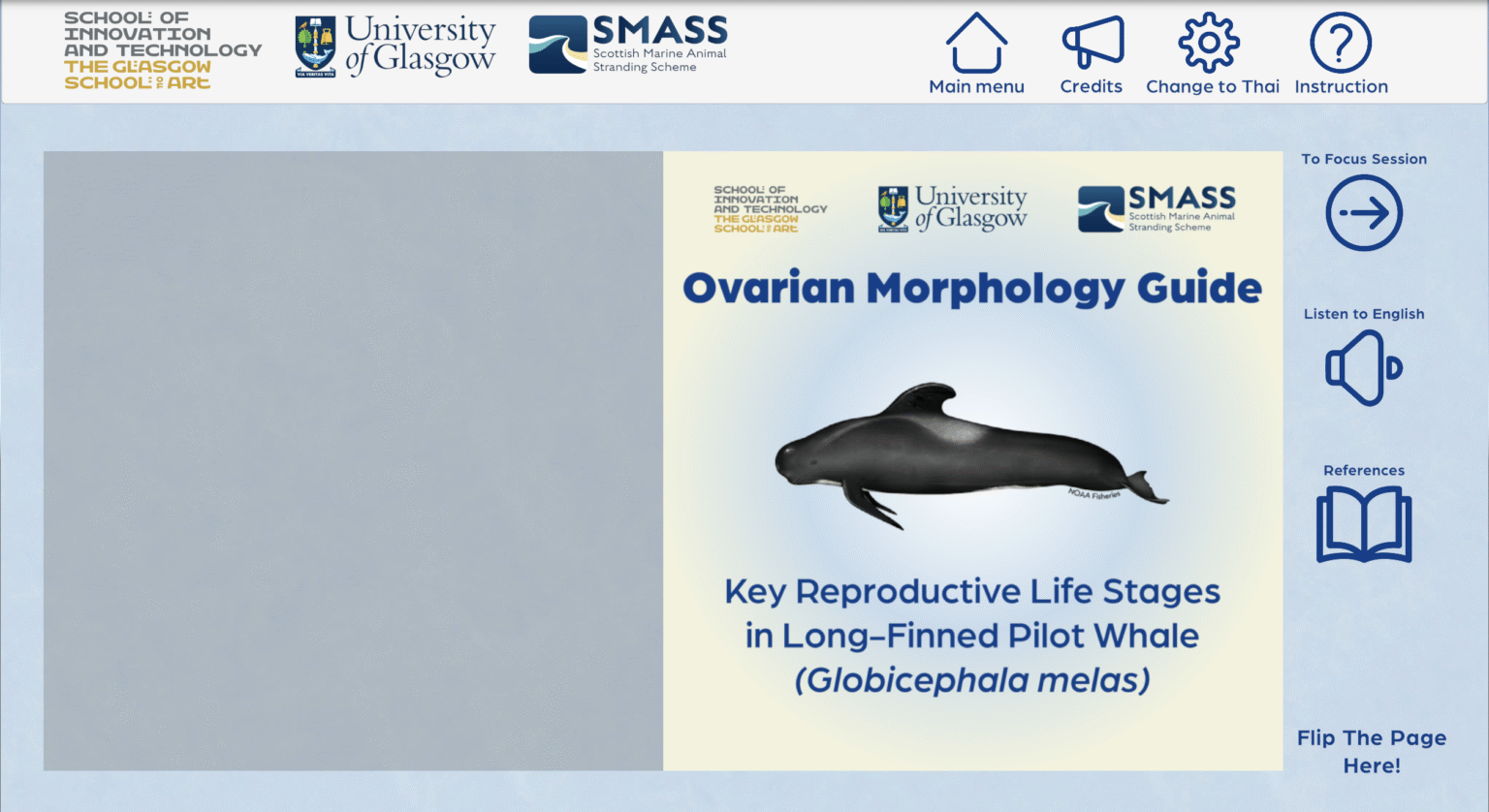
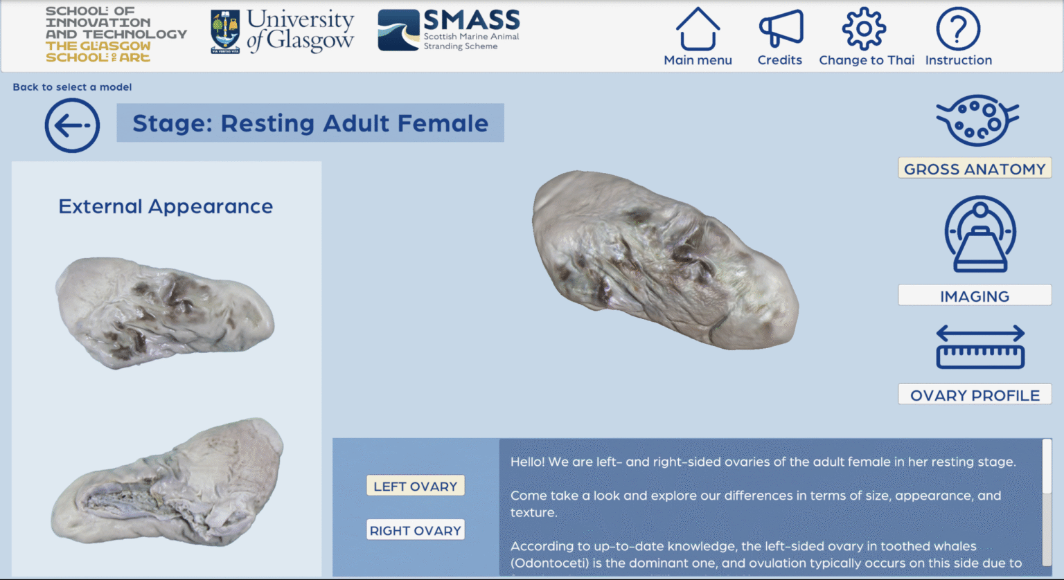
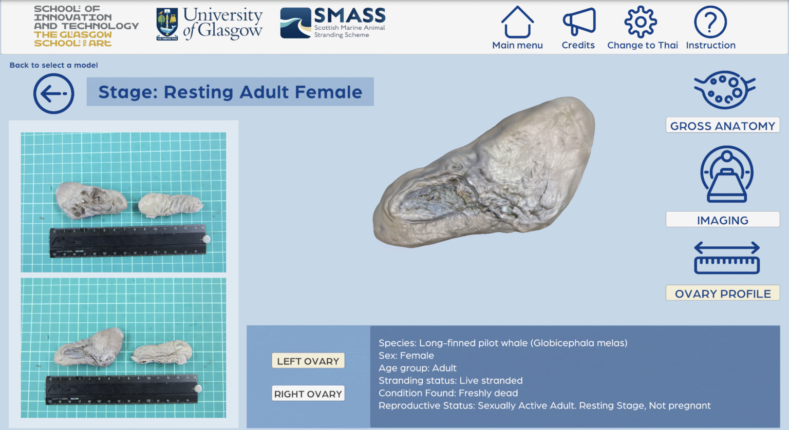
This project explored the effectiveness of an interactive 3D webGL-based learning tool for visualising ovarian transformation in different reproductive stages in female long-finned pilot whales (Globicephala melas).
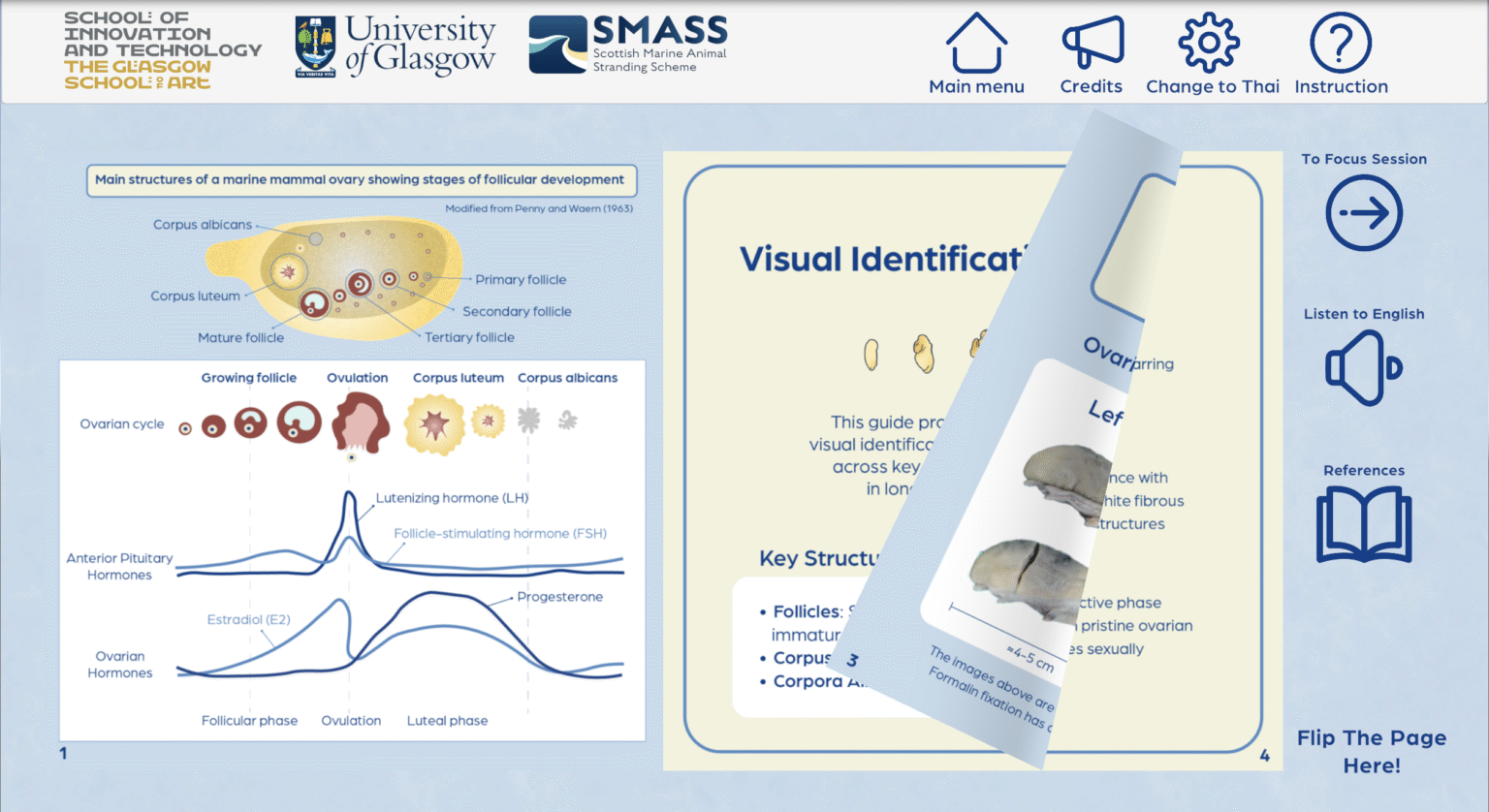
Knowledge gaps in the female reproductive system in cetaceans are limited due to limited access to specimens, the feasibility of encountering animals in the wild, and reliance on conventional learning methods such as histology and static textbook images. These reduce the possibilities of understanding three-dimensional complexity to a two-dimensional representation.
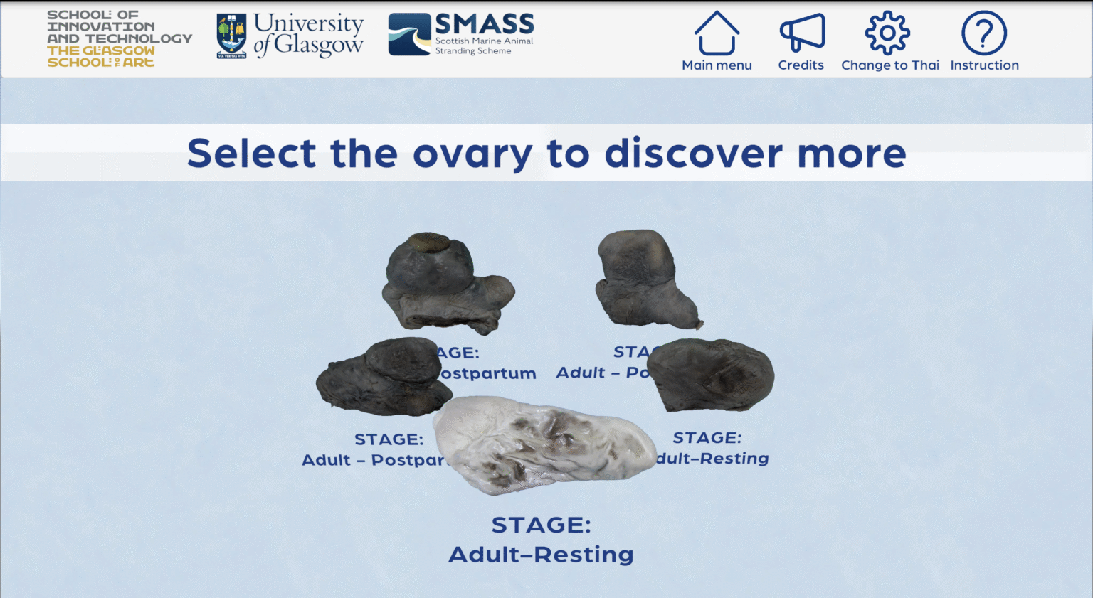
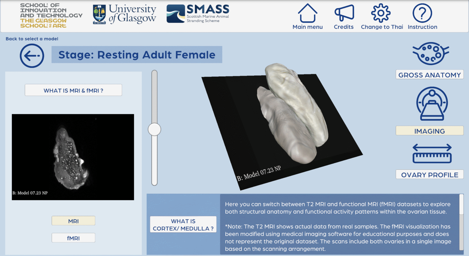
This research developed and evaluated an interactive 3D WebGL-based learning platform that transforms MRI data to visualise ovarian anatomical transformations across key reproductive life stages: adult female in the resting stage and post-partum female. The application was assessed with veterinary professionals, marine mammal researchers, and others from related fields using five evaluation metrics: knowledge assessment, cognitive load, face and content validity, usability, and user motivation (RIMMS).
Overall, the results presented significant improvement in understanding and positive perceptions towards the application, demonstrating the potential of 3D visualisation to enhance anatomical understanding in marine mammal science. It also provides a meaningful direction for interdisciplinary communication among professionals and opens doors for more research toward medical visualisation in veterinary science.
Project Links
3D Modelling and Animation by Orn
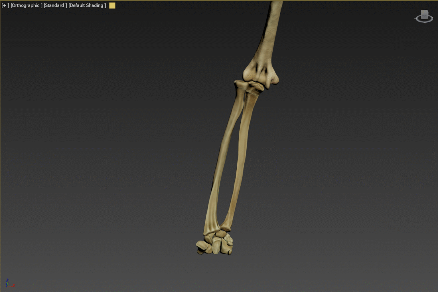
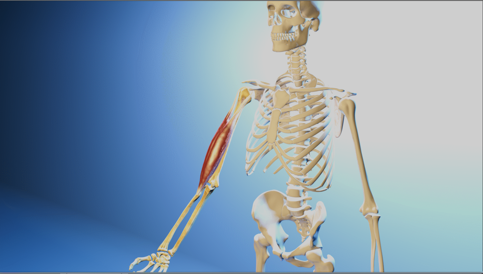
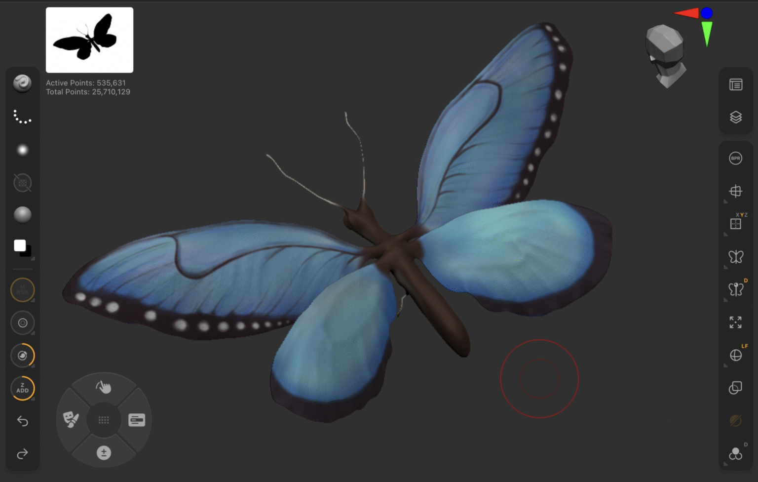
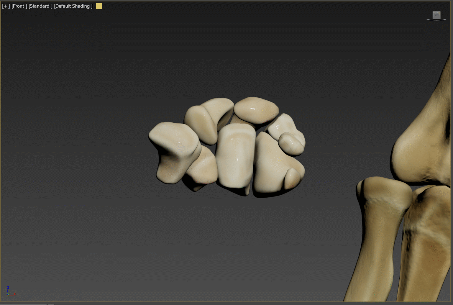
Part of Human Anatomy; Human Skeleton
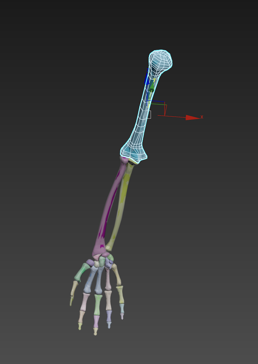
This project was developed as part of a 3D modelling course, where I created detailed anatomical models of the human upper limb. The collection includes the humerus, radius, ulna, carpals, metacarpals, and phalanges, complemented by a bicep muscle model. The animation aims to present the muscular movement of the bicep and the motion of the blue morpho butterfly.
Project Links
Volumetric Visualisation by Orn
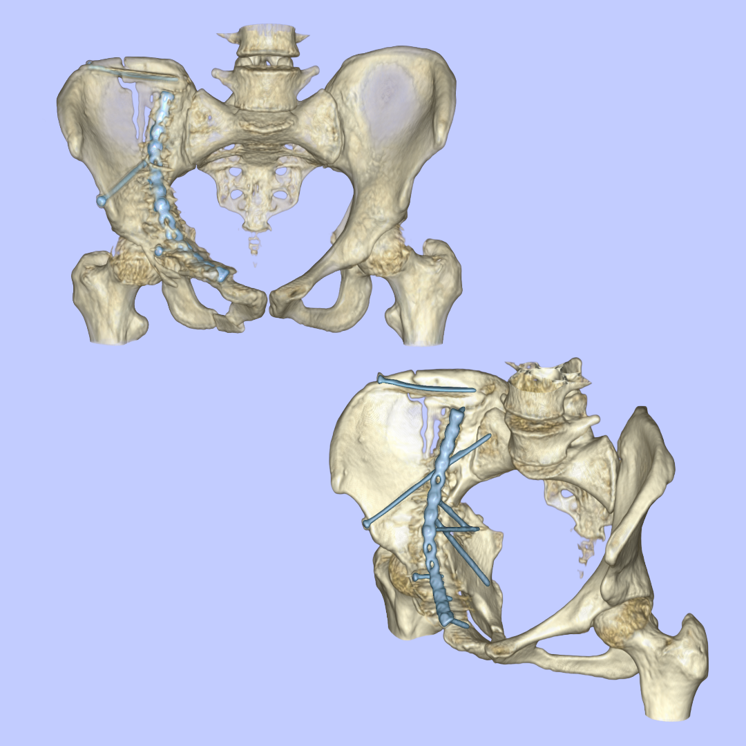
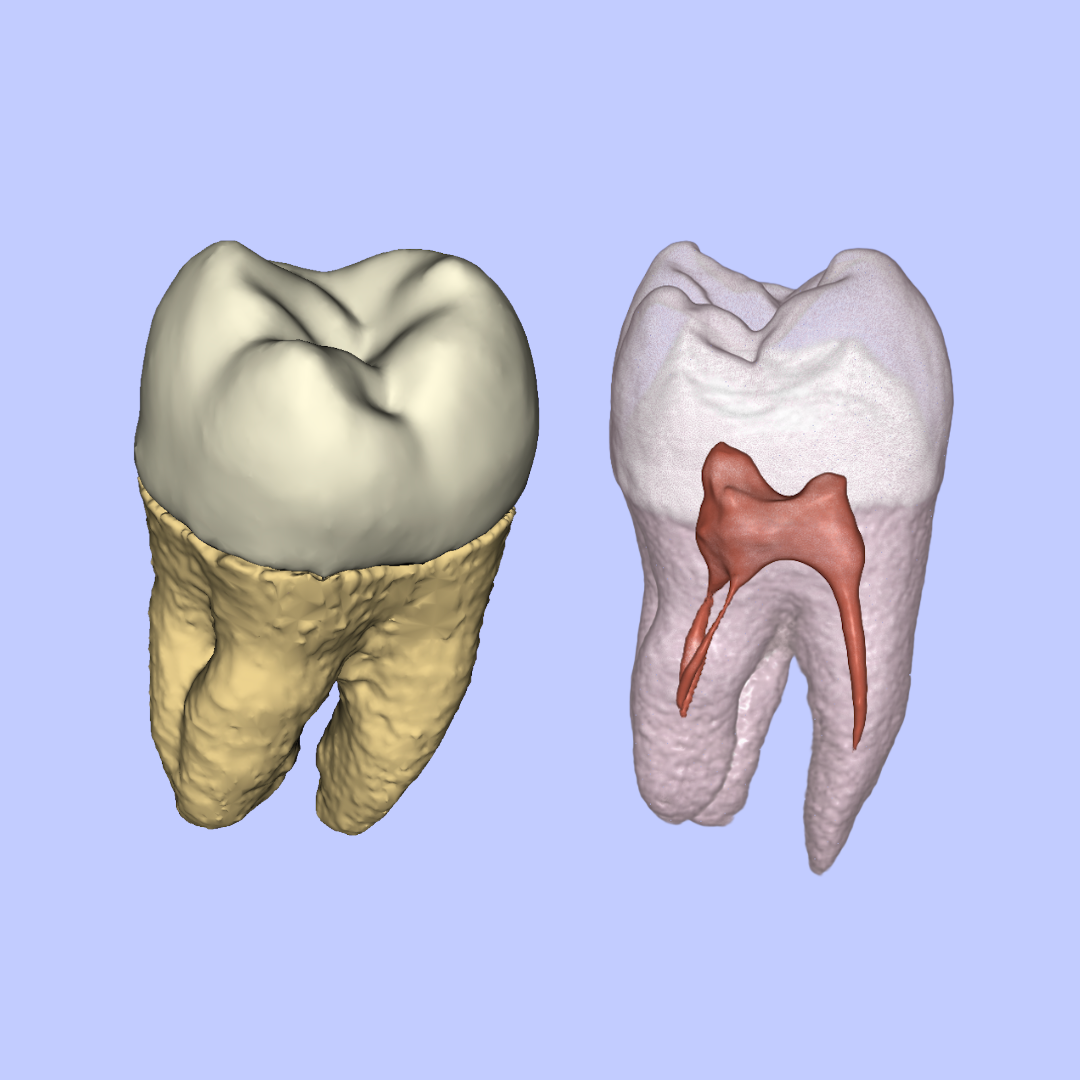
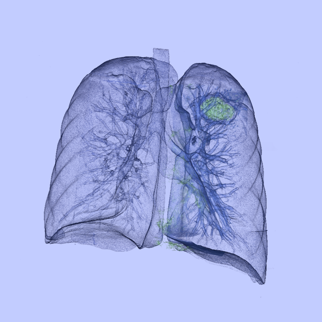
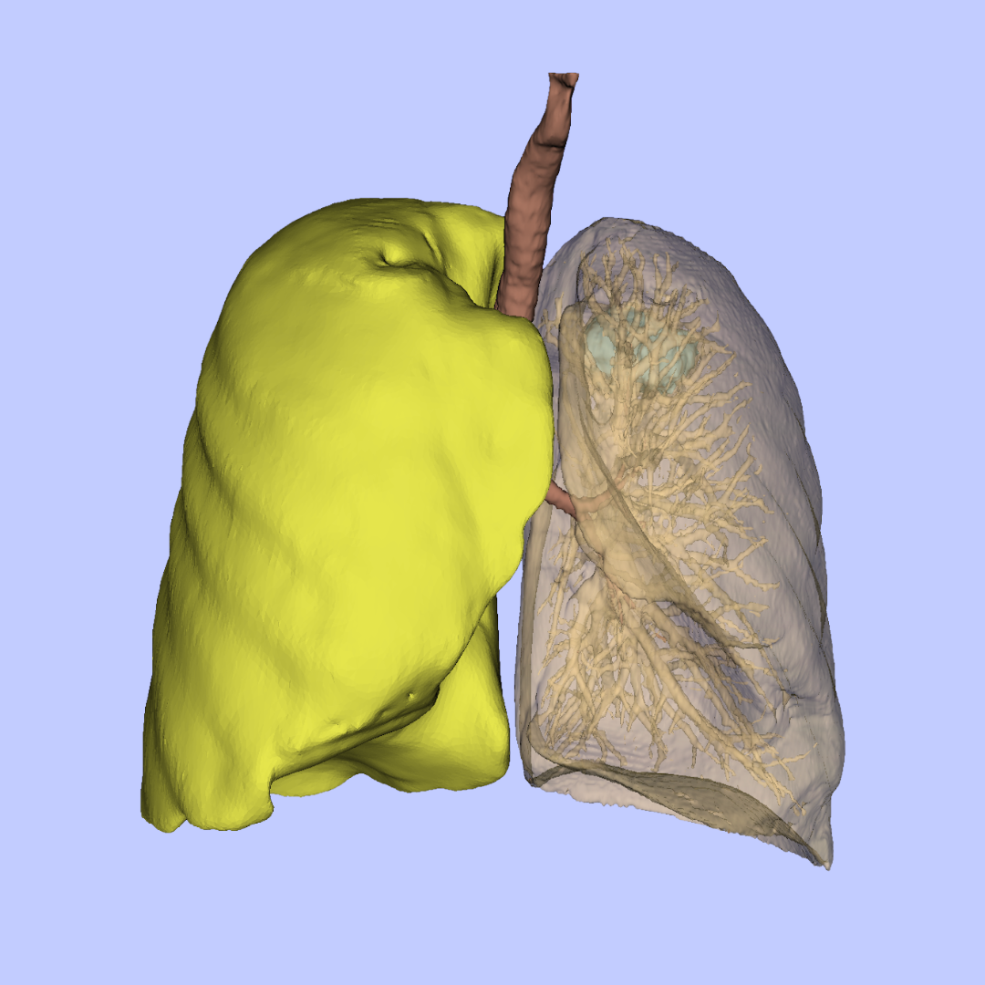
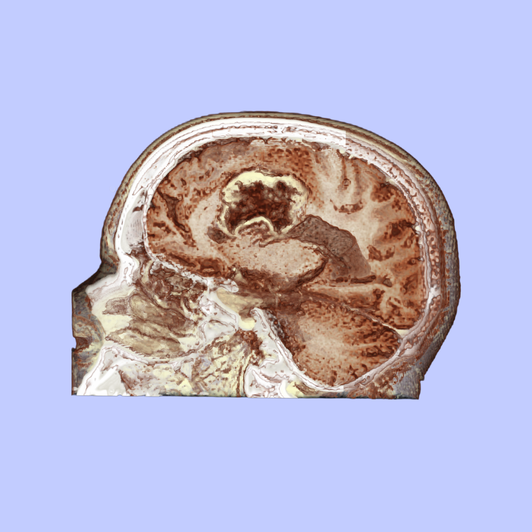
Human anatomy, Medical imagings
Volumetric visualisation is a powerful technique that transforms stacks of medical imaging data, such as MRI and CT scans, into three-dimensional models. Using specialised software like 3D Slicer or MITK, I can reconstruct these datasets and manipulate their colour, contrast, and transparency to reveal anatomical structures from new perspectives. This process transforms flat, two-dimensional medical images into 3D representations that enhance the understanding and communication of complex anatomical relationships, both spatial and temporal.
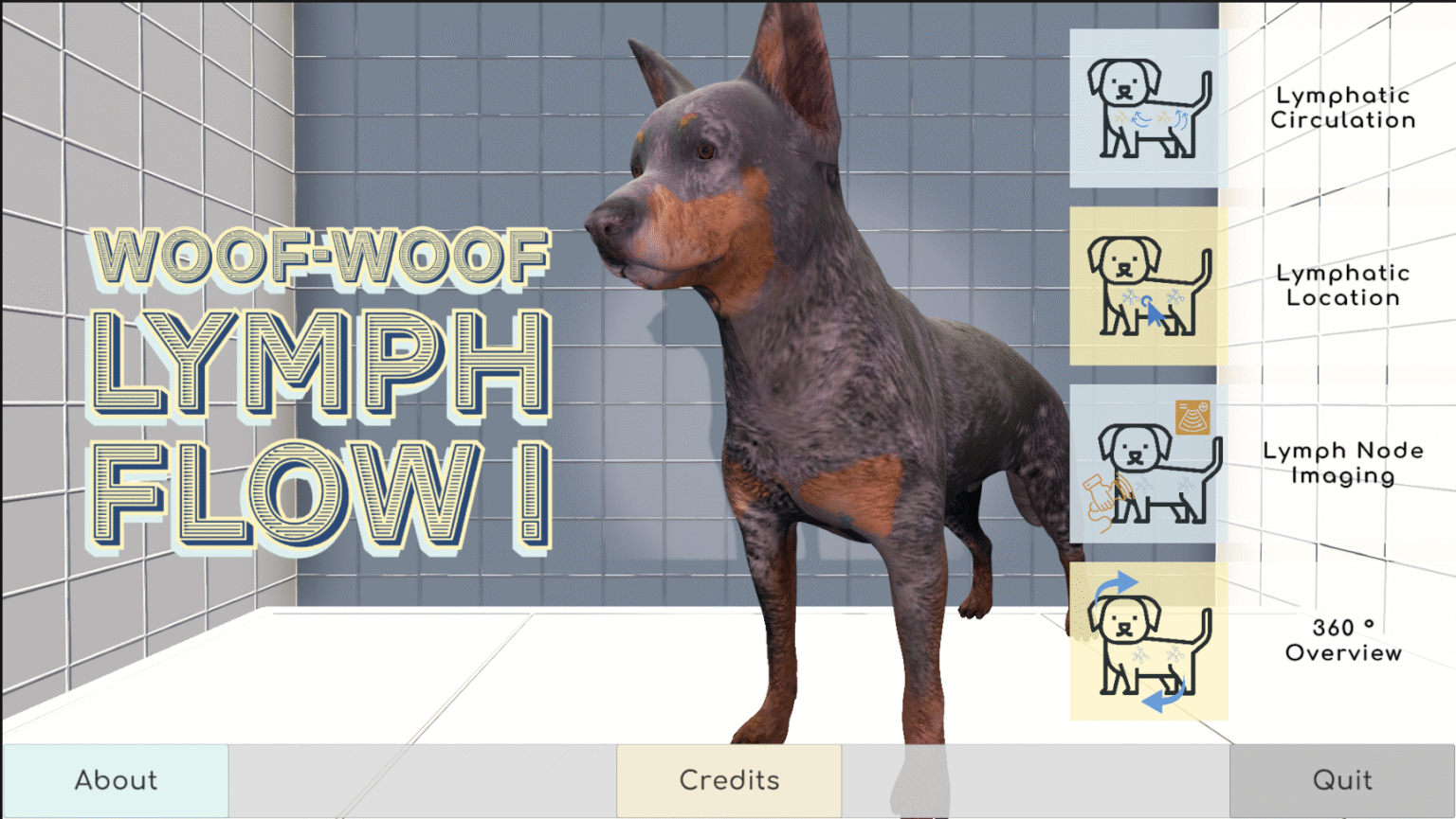
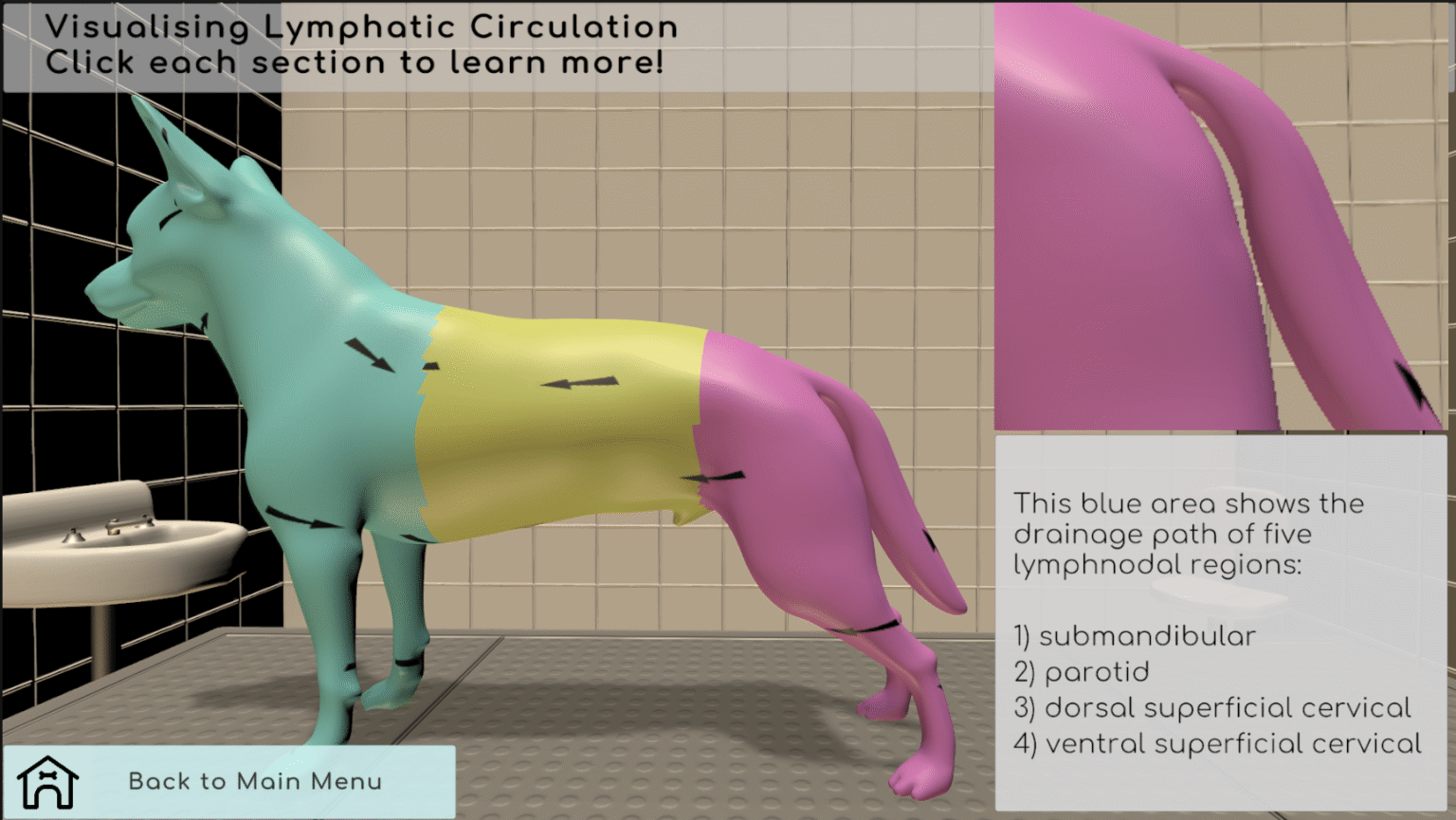
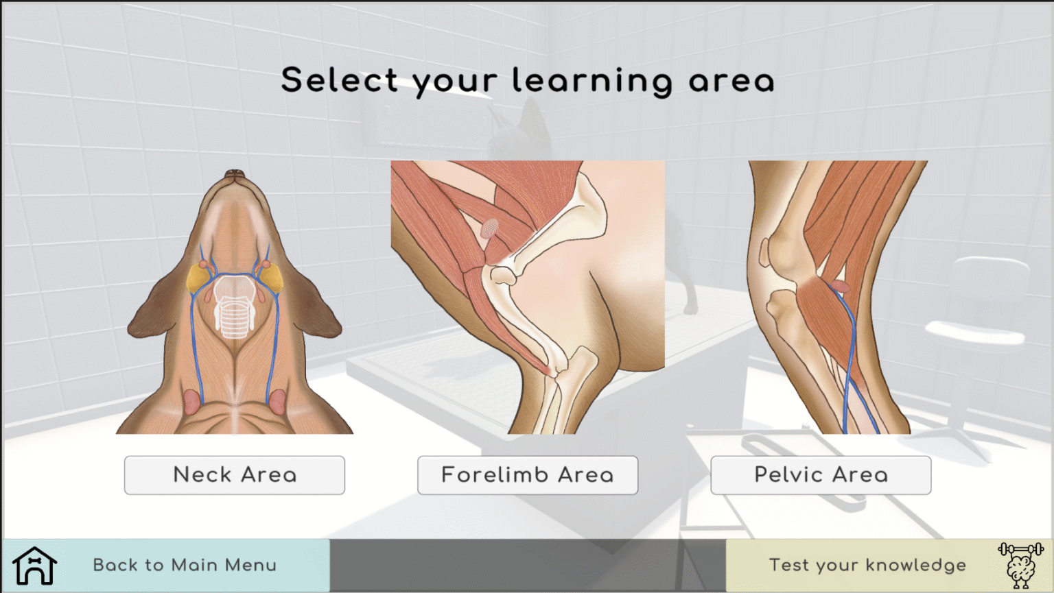
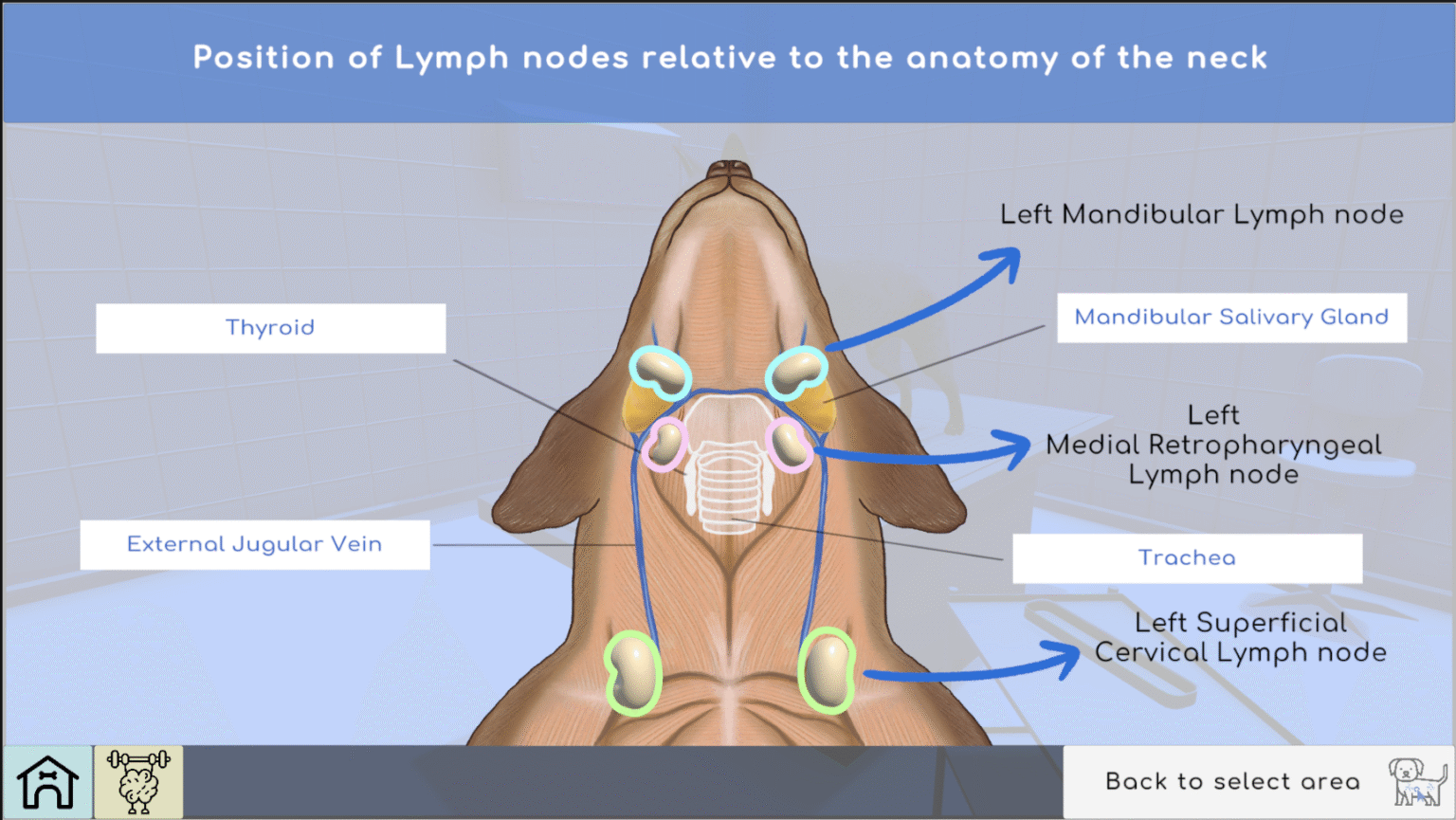
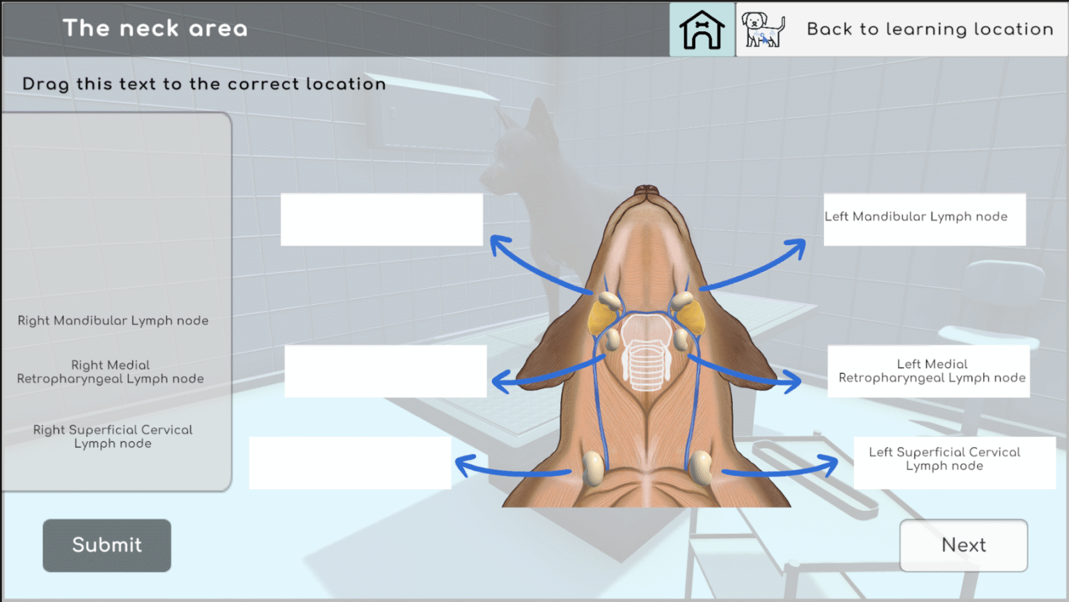
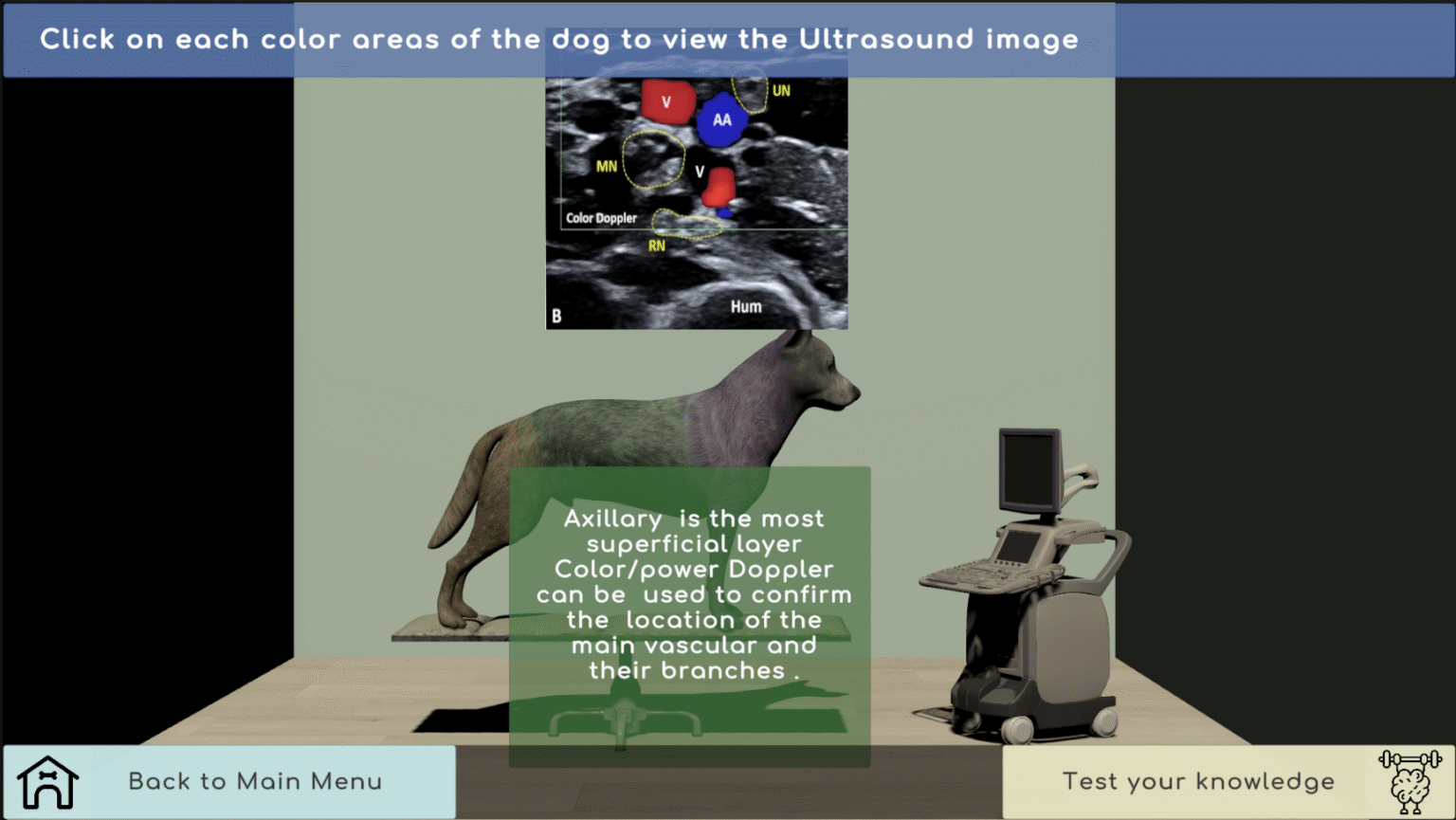
WoofWoof LymphFlow
Developed in Unity, this interactive application makes learning canine lymphatic anatomy engaging and accessible. Users can explore lymph nodes by region, test their knowledge with drag-and-drop exercises, observe lymphatic flow visualisation, and examine related ultrasound data. This project created a comprehensive educational experience that transforms traditional anatomy study into active learning. This group work was developed as part of my interactive application coursework at GSA.