MSc Medical Visualisation & Human Anatomy School of Innovation & Technology
Rebecca Millar

Hello! My name is Rebecca Millar and I’ve always been interested in how to visualise anatomy in 3D, because I’ve always found learning from 2D images quite difficult. That curiosity started during my BSc (Hons) in Neuroscience at the University of Glasgow, where I developed a strong interest in medical imaging, neuroanatomy, and finding easier ways to understand complex structures.
Wanting to explore this further, I pursued a Master’s in Medical Visualisation and Human Anatomy, where I’ve developed skills in volumetric visualisation, 3D modelling, Unity app development, and coding in C#. I’ve also had the rare opportunity to gain hands-on experience through cadaveric dissection, allowing me to connect my digital work with real-world anatomy.
My thesis project: an MRI-based atlas of the female pelvic region, has strengthened my ability to work with imaging data and translate it into 3D, interactive, user-friendly visual tools. Looking ahead, I’m excited to apply my technical skills, anatomical knowledge, and passion for 3D visualisation to create innovative solutions that make anatomy clearer, more accessible, and ultimately improve patient care.
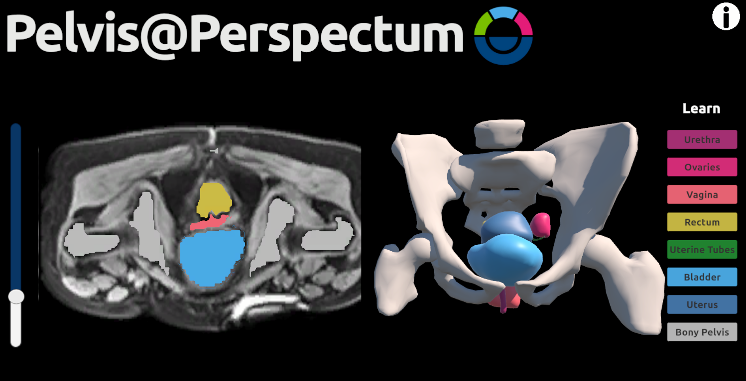
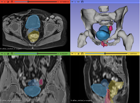
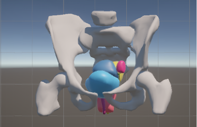
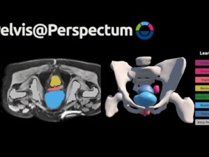
Pelvis@Perspectum – An MRI Atlas of the Female Pelvic Region
My thesis project was conducted in collaboration with Perspectum, a medical technology company specialising in advanced imaging and data-driven healthcare solutions. The project was developed in response to a key challenge in anatomical education: the difficulty learners face in conceptualising the complex spatial relationships of female pelvic anatomy when relying solely on traditional two-dimensional resources such as static MRI slices or textbook diagrams.
Three-dimensional reconstructions of anatomical structures are increasingly recognised as valuable tools for education, training, and clinical communication. When combined with MRI data, they have the potential to bridge the gap between abstract anatomical concepts and their clinical context, enabling more intuitive understanding. This is particularly relevant in pelvic anatomy, where the spatial arrangement of organs is both intricate and highly variable.
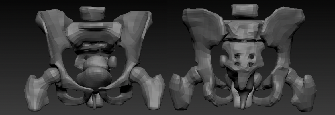
The aim of this project was to create anatomically accurate 3D models of female pelvic organs from high-resolution MRI data and integrate them into an interactive web-based application designed to support anatomical learning. MRI data provided by Perspectum was manually segmented in 3D Slicer to produce anatomically accudate models, which were then optimised in ZBrush for clarity and performance. The final models were incorporated into a Unity application, allowing users to compare axial MRI slices with their corresponding segmented 3D structures in real time, with features such as structure toggling, colour-coded segmentation, and interactive rotation.
This work demonstrates the potential of MRI-based 3D visualisations to enhance anatomical understanding and lays the groundwork for future developments.
I would like to thank my supervisors, Dr. Matthieu Poyade, Dr. Caroline Allen, and Ella Jones for their continuous support and guidance throughout this project.
3D Modelling and Animation
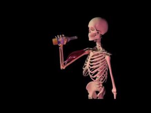
For the 3D Modelling and Animation module, I created a project called “Anatomy of Having a Beer” using 3DS Max and ZBrush.
The project gave me hands-on experience with the full 3D modelling workflow, including modelling, texturing, and rendering. I also explored aspects of animation to bring the model to life. What I enjoyed most was seeing how different skills, from anatomy to technical problem-solving, come together to create a cohesive and visually engaging final piece.