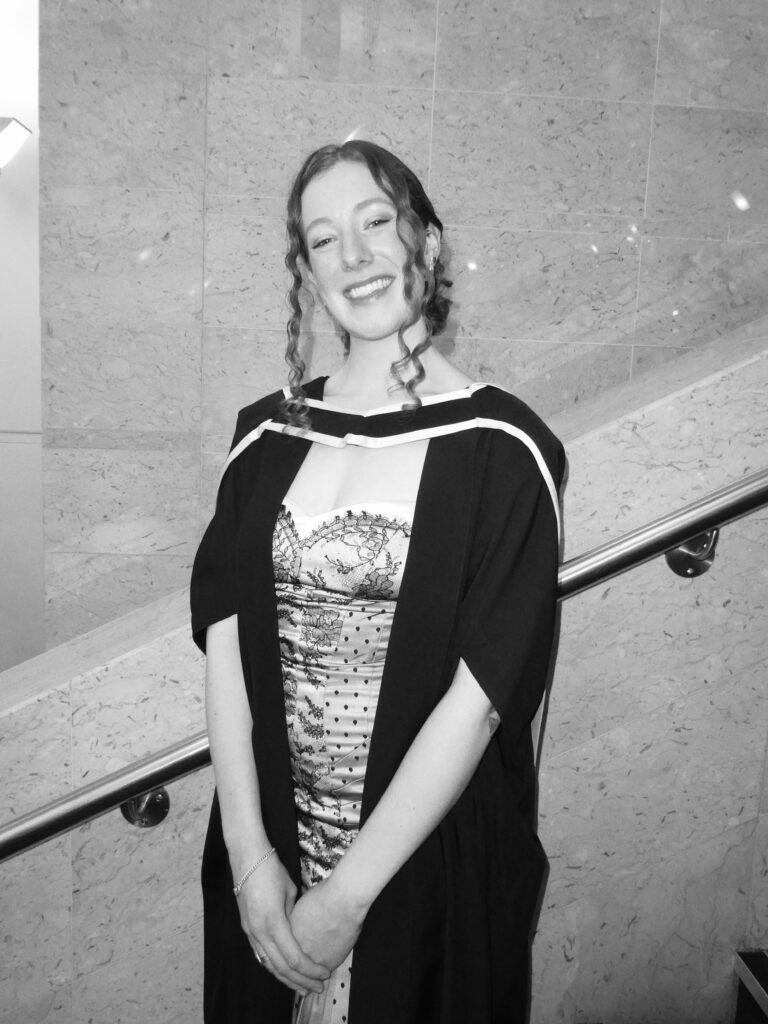
MSc Medical Visualisation & Human Anatomy School of Innovation & Technology
Kate MacDiarmid
I hail from Calgary, Alberta, Canada and completed my undergraduate degree in Neuroscience in 2021 at Dalhousie University (Halifax, Nova Scotia). Prior to undertaking the programme, I spent a couple of years indulging my passion for travel, embarking on working holiday visas in both Australia and New Zealand and backpacking in Portugal, Morocco, and Indonesia.
I was inspired to join the Medical Visualisation programme due to its unique intersection of creative thinking, health sciences, and cutting edge technology. The programme has provided me with a range of industry-standard skills in 3D modelling, animation, interactive app design and development, and 3D printing. I am excited at the prospect of being able to apply these skills as I embark on my career here in the UK.
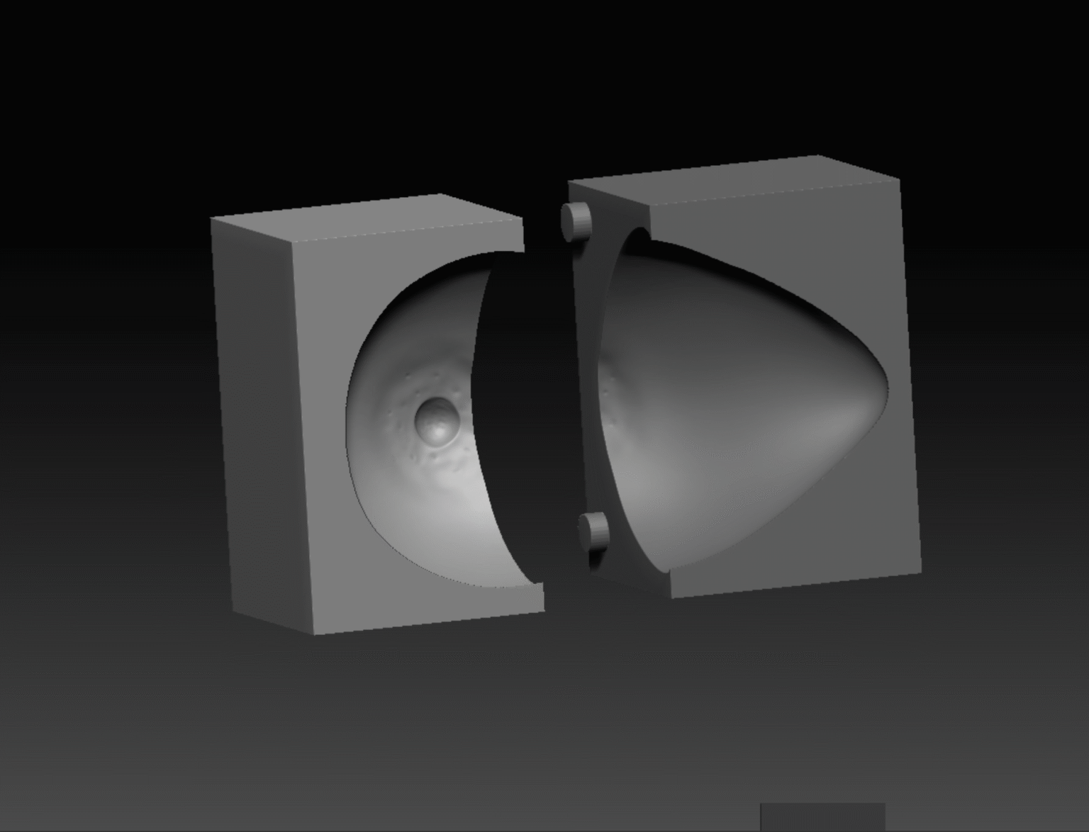
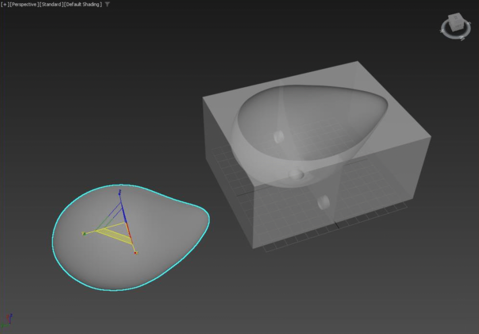
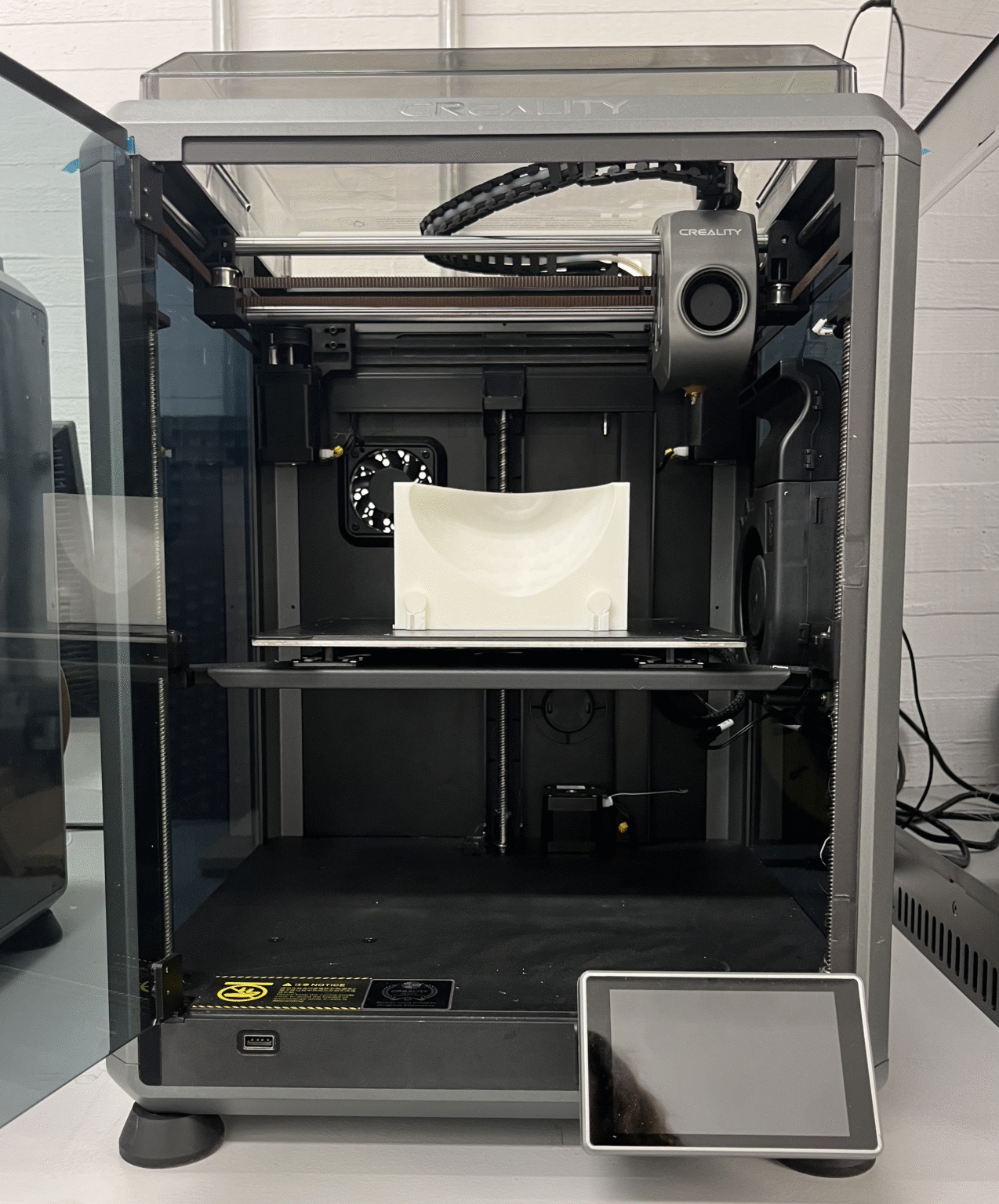
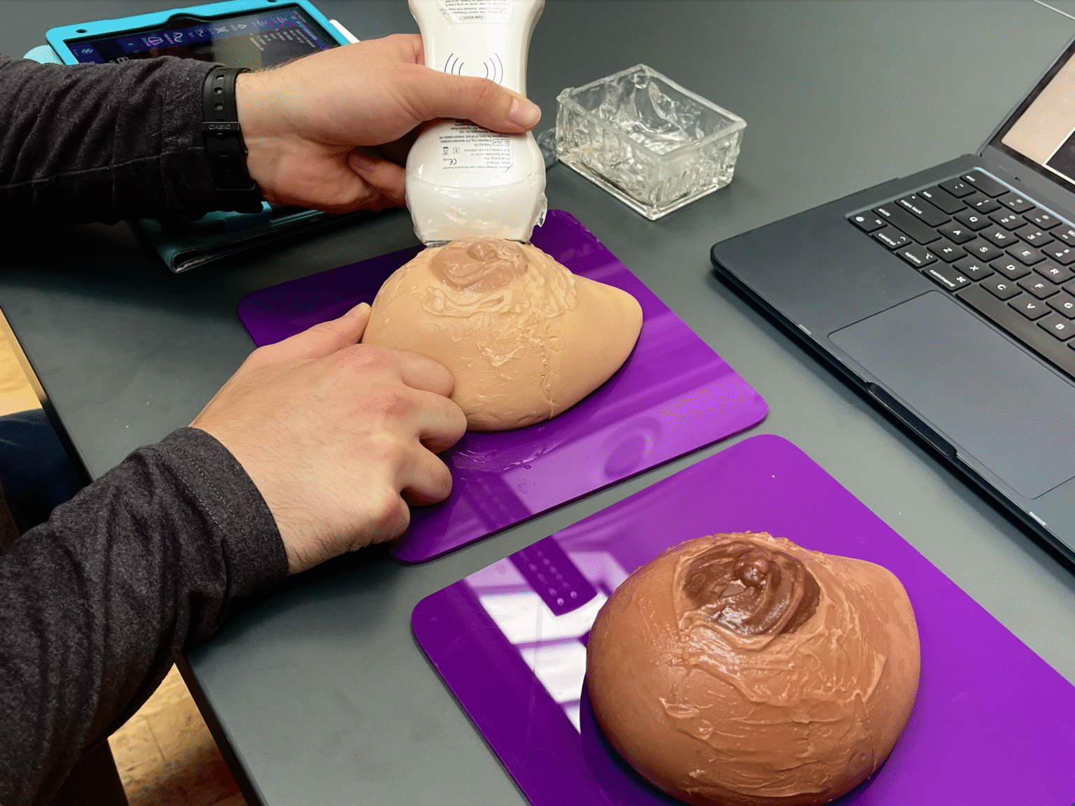
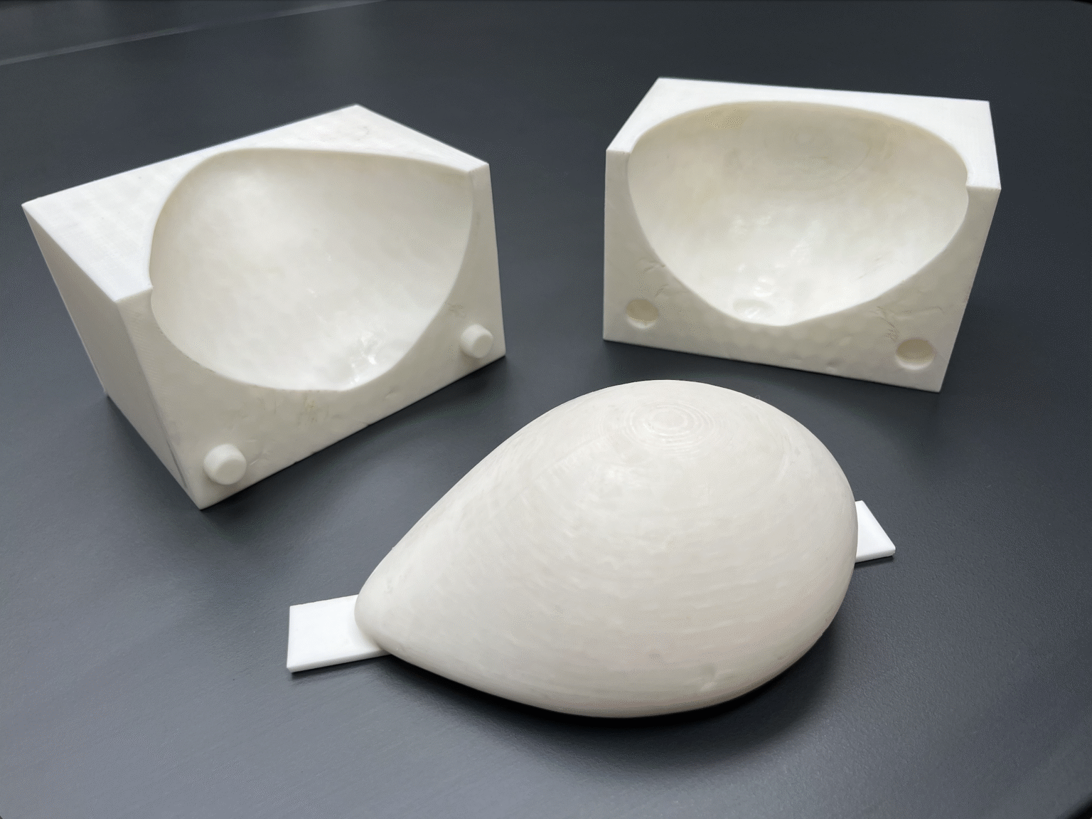
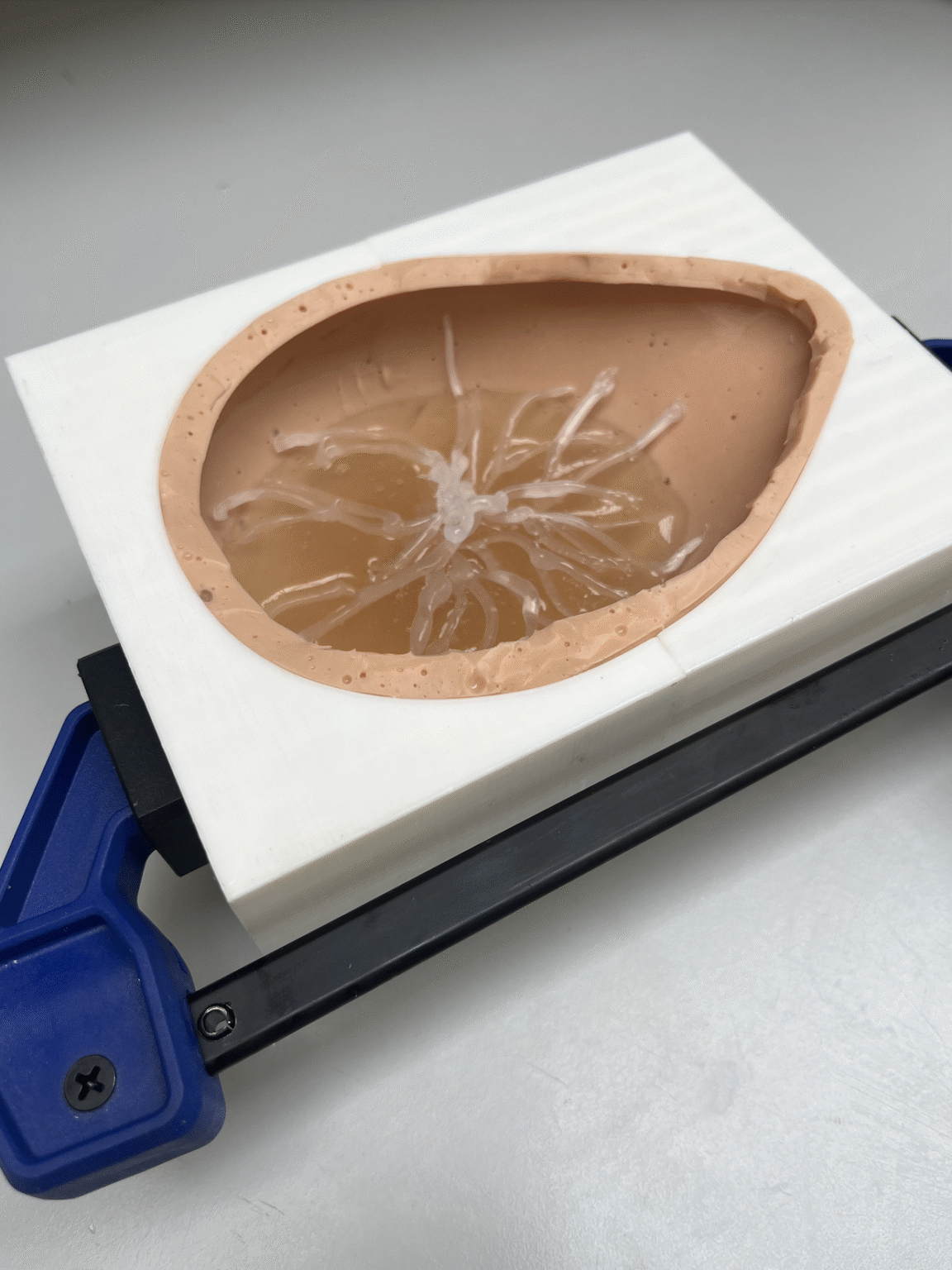
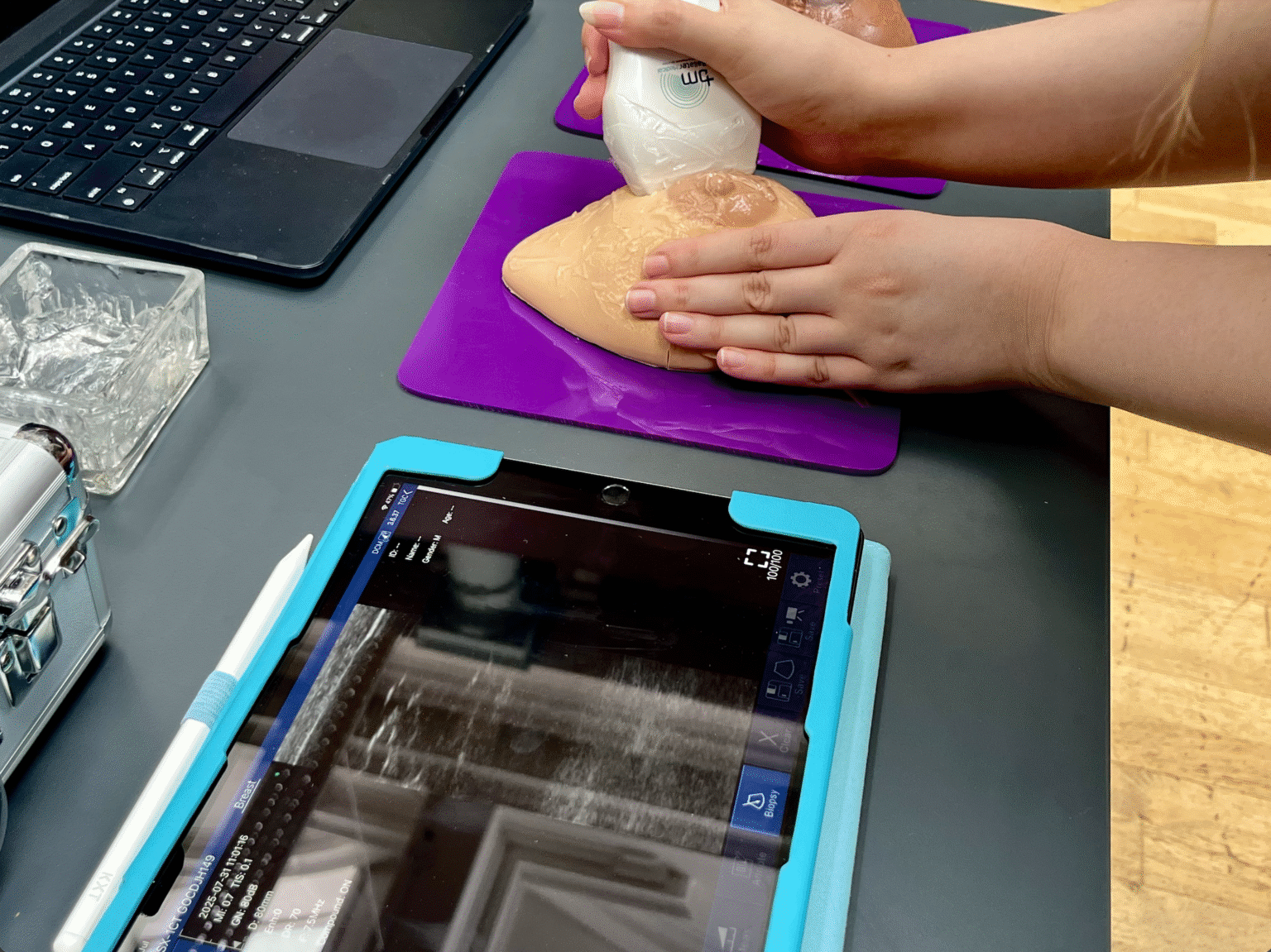
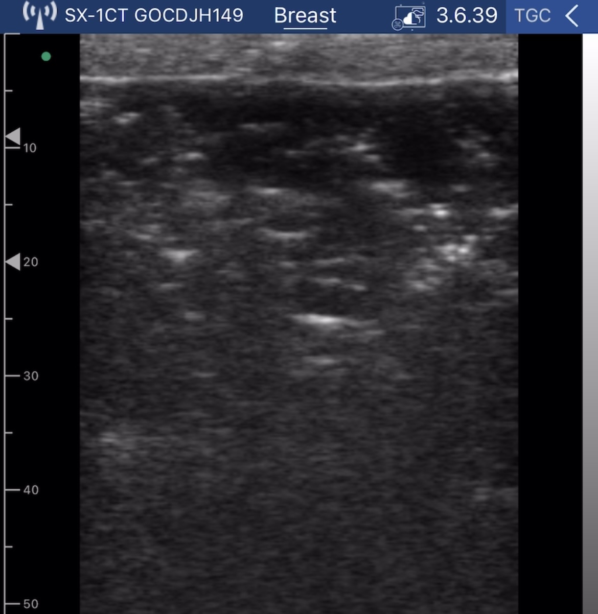
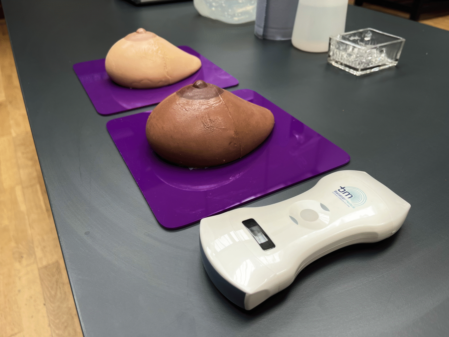
The development of a realistic, adjustable-density breast phantom with 3D printing for use as an ultrasound training tool
This work was my thesis project for the Medical Visualisation and Human Anatomy programme. The aim of the project was to create realistic-looking breast tissue training simulations (“phantoms”) with diverse skin tones that also demonstrated different levels of breast density under ultrasound. Two phantoms were developed, one in a light-medium skin tone with a higher density, and one with a dark skin tone and lower density. Only low-cost materials and basic equipment were used in construction, making creation and distribution of the phantoms accessible. The phantoms were critically evaluated and contrasted by participants with various life science backgrounds, all of whom found the phantoms to be useful training tools and the presence of diverse skin tones to be an important addition.
This project allowed me to combine my interests in women’s health, diversification of healthcare educational materials, and healthcare accessibility. While some tweaks to the design are required, there is potential for future development and commercialisation of these breast phantoms into a low-cost alternative to those currently on the market.
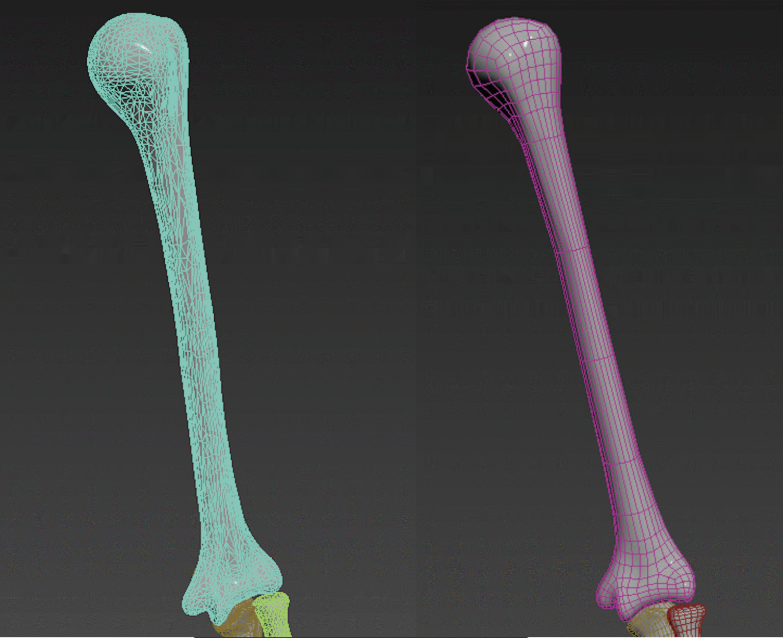
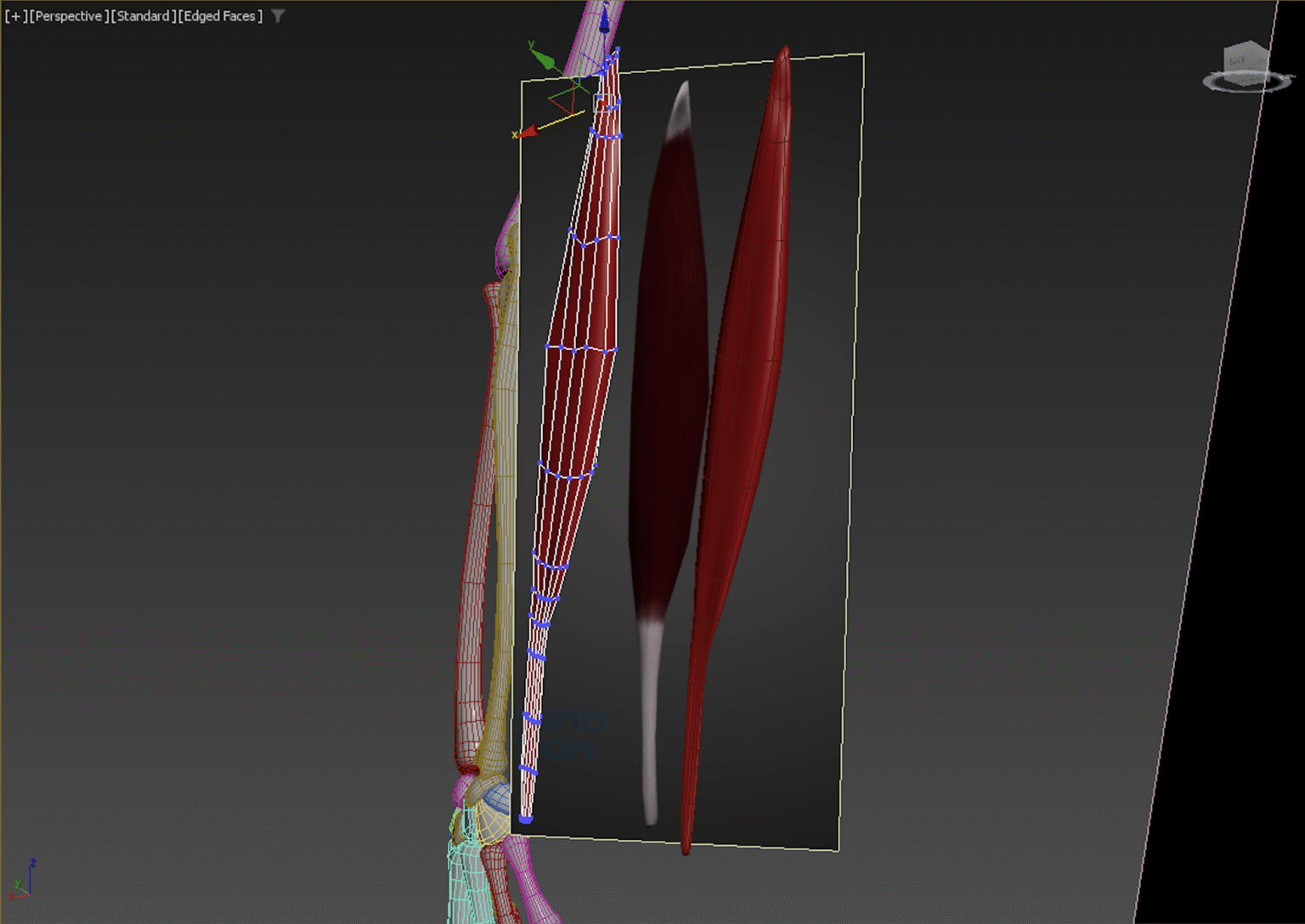
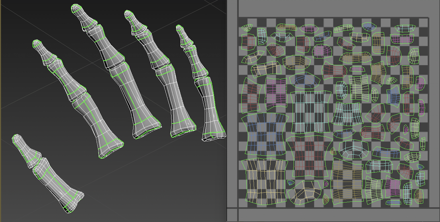
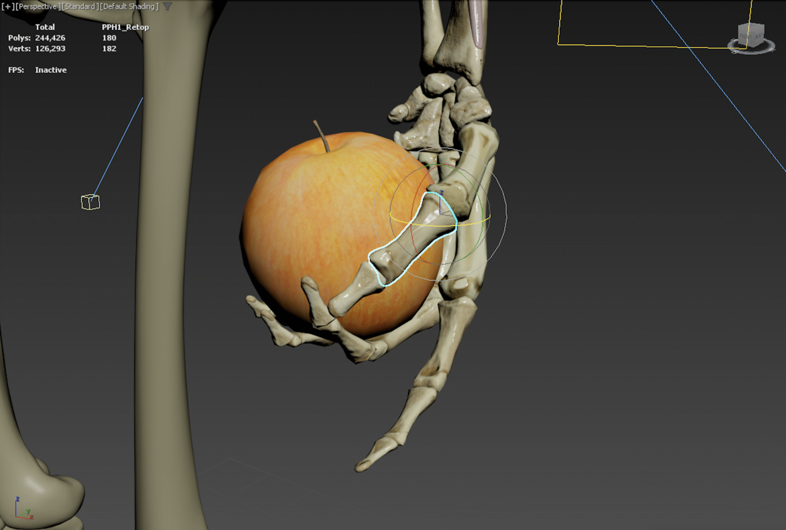
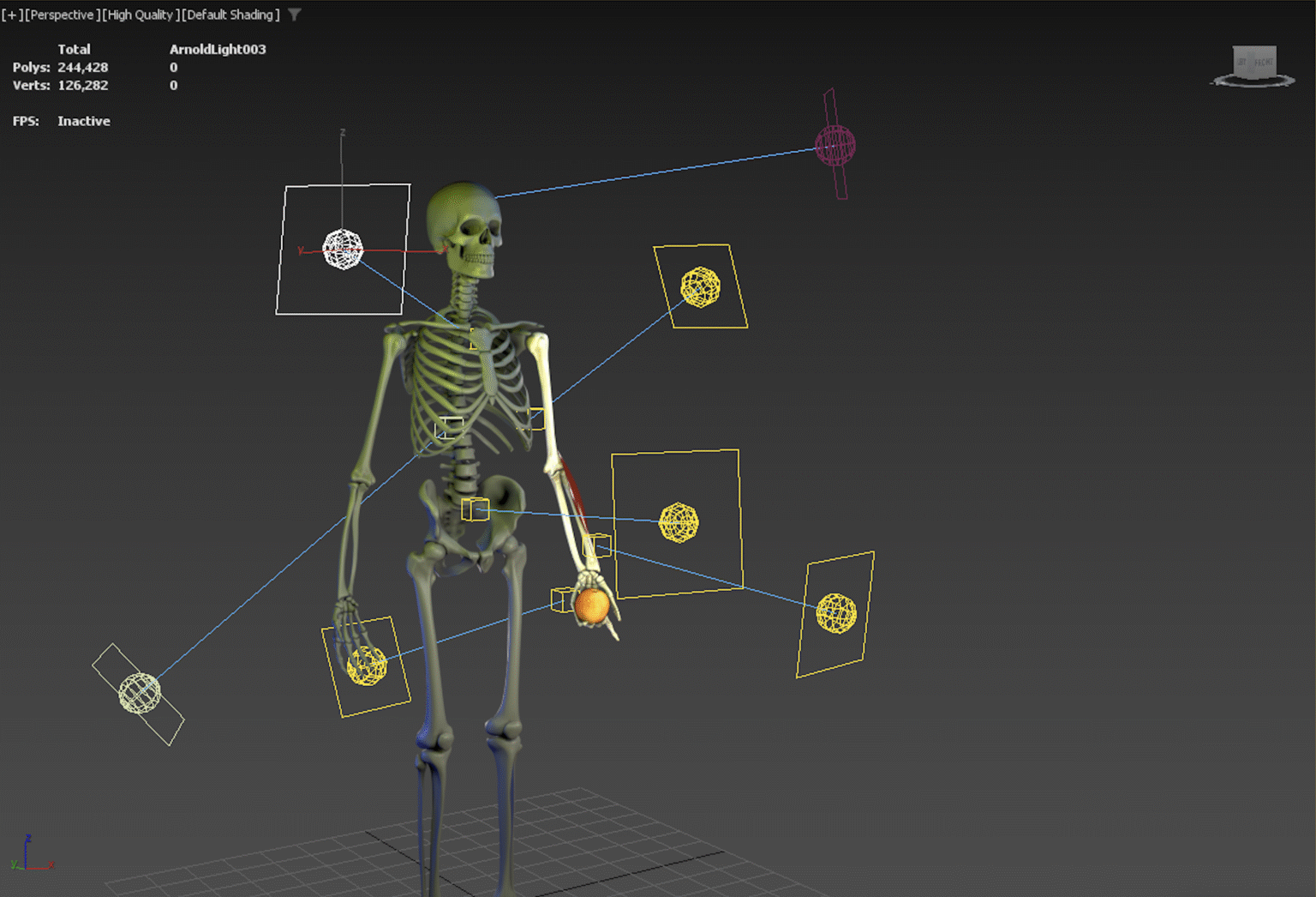
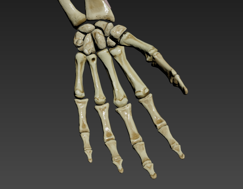
3D modelling
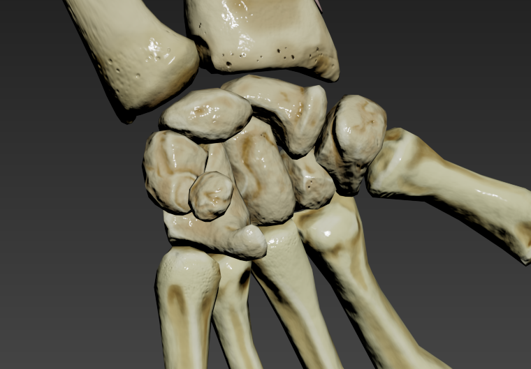
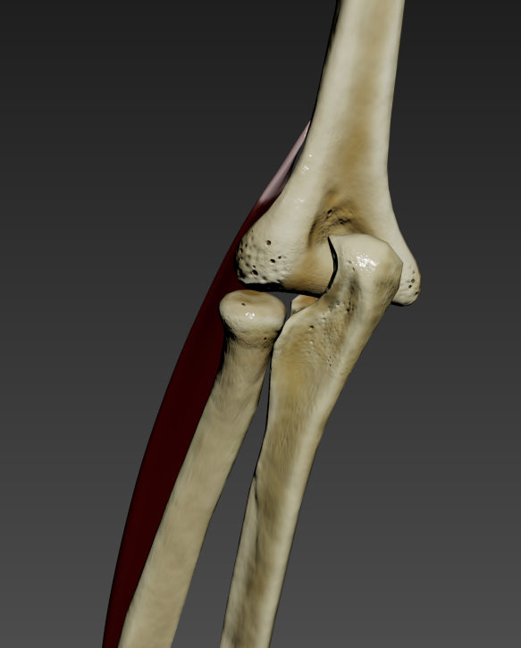
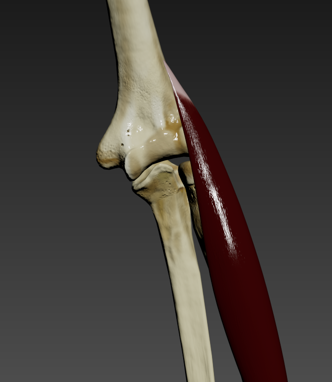
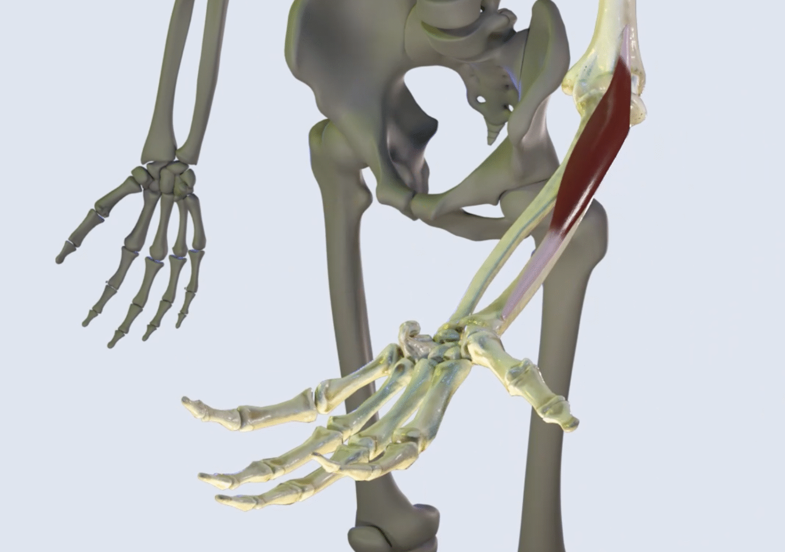
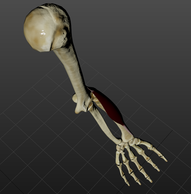
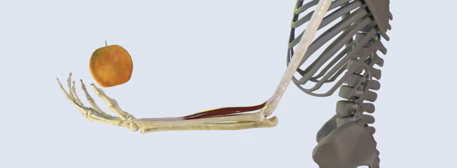
This 3D animation of the action of brachioradialis was completed using ZBrush and 3ds Max, leading industry-standard modelling and rendering software. Brachioradialis is a small muscle in the forearm, the action of which is demonstrated by executing a throwing motion. All 30 bones of the upper limb were individually retopologized, sculpted, and textured from primitive assets, and the muscle was box modelled from scratch. This project was highly beneficial for gaining an understanding of the variability and flexibility of workflows that can be undertaken to create such a render. Realistically animating the effect of gravity on the apple and the motion of the outstretched hand was a unique challenge in this assignment.
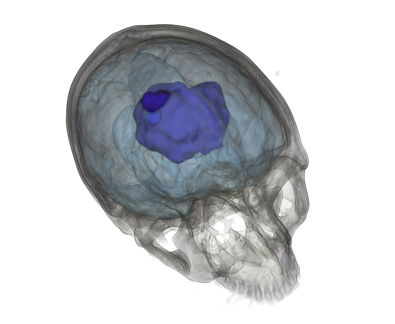
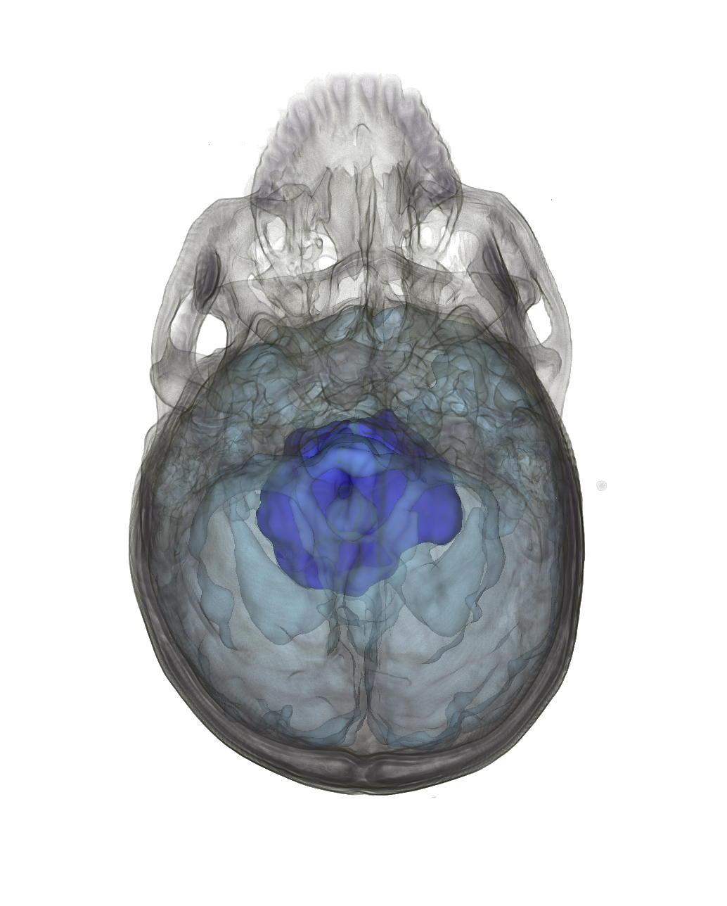
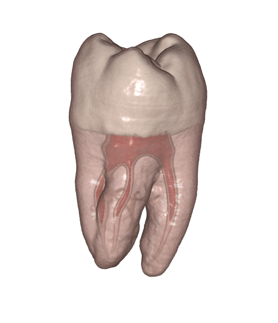
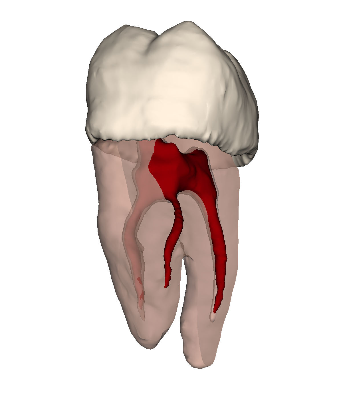
Volumetric visualisation
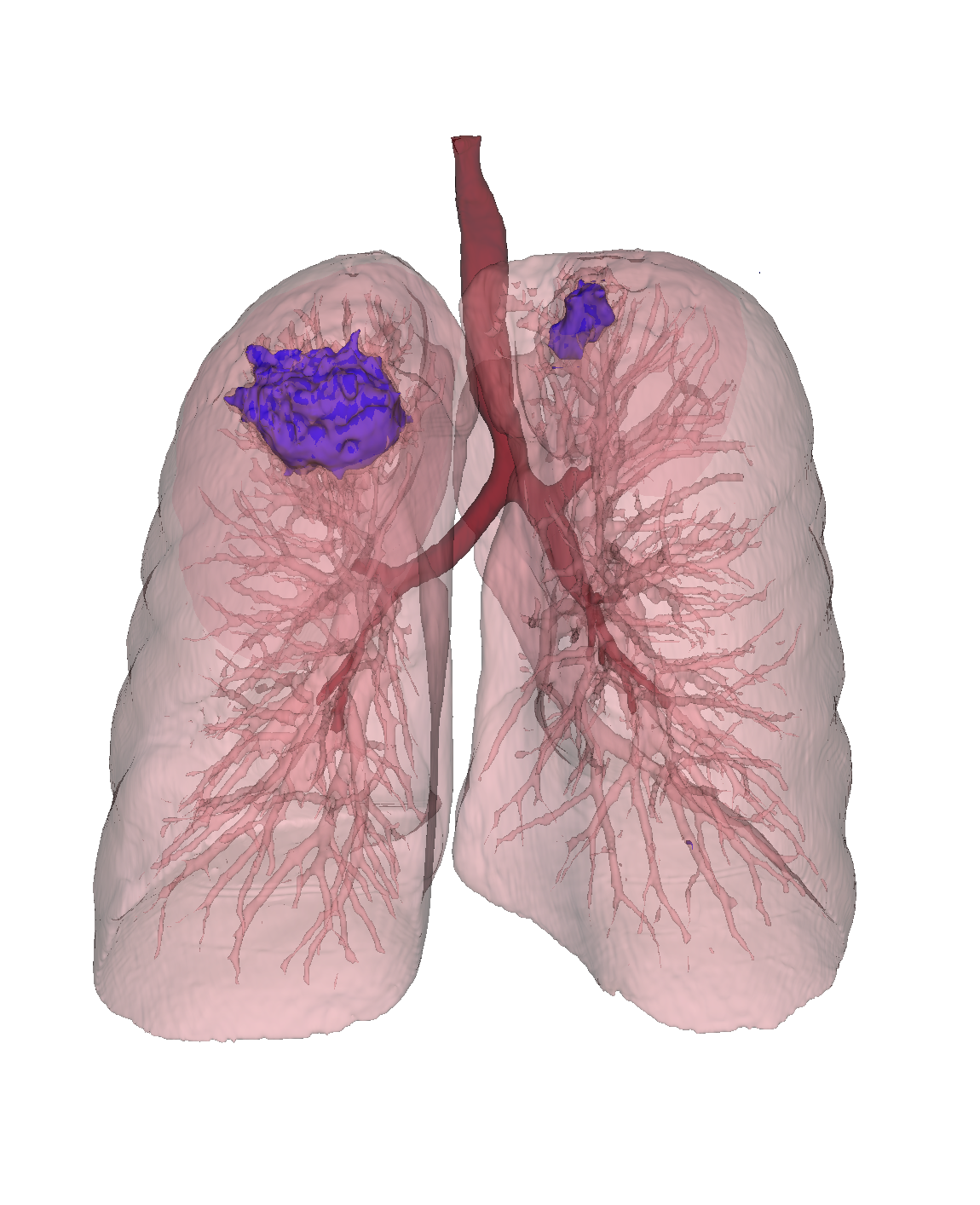
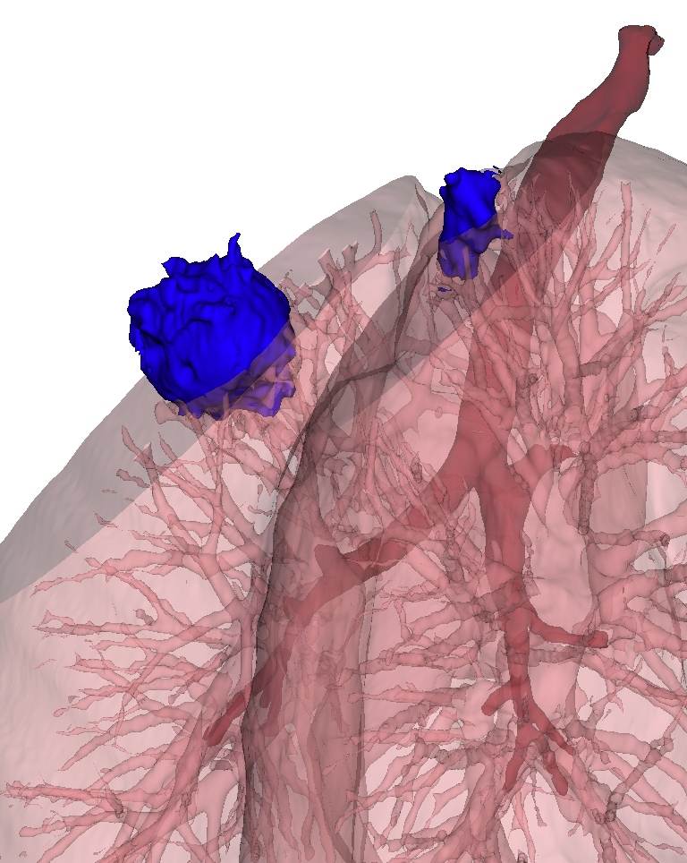
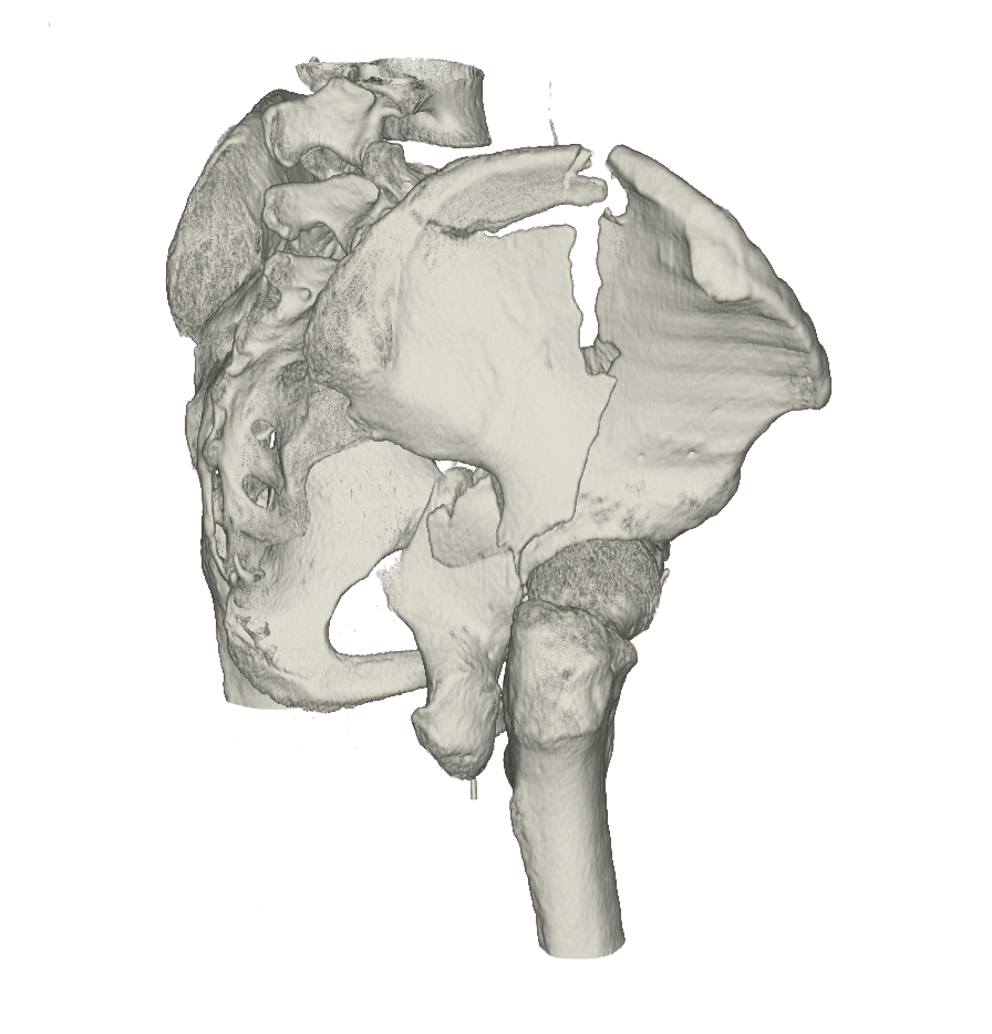
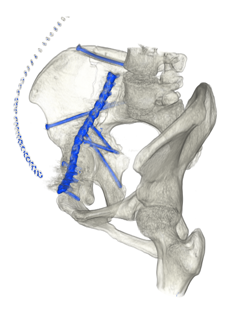
This work comprises a series of exercises completed for the Volumetric Visualisation course, where 3D models were constructed from medical imaging datasets. Both direct and indirect volume rendering techniques were used. I greatly enjoyed the challenges of segmenting anatomical areas from different types of datasets and developing creative solutions for displaying the data in the best manner. It was intriguing to see the ways in which my classmates visualised the same datasets differently. I look forward to continuing to grow my skills in the software used in this course.
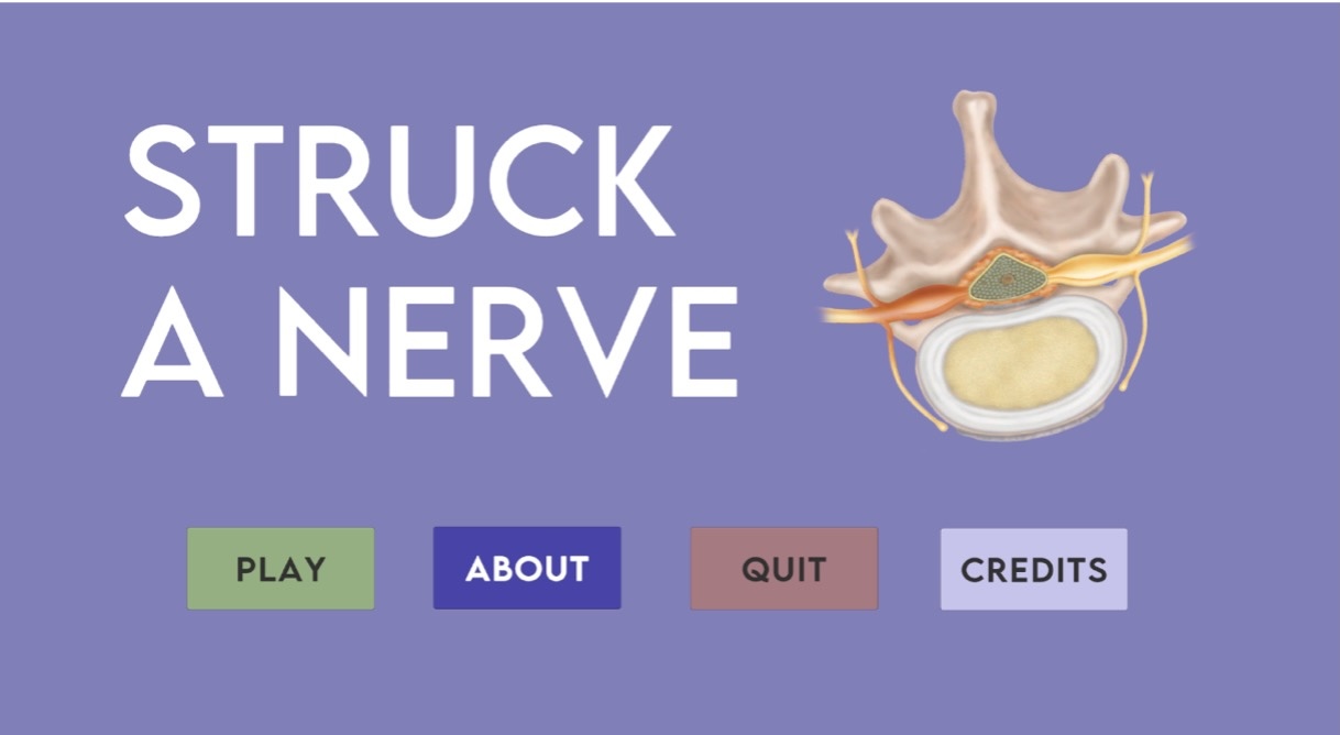
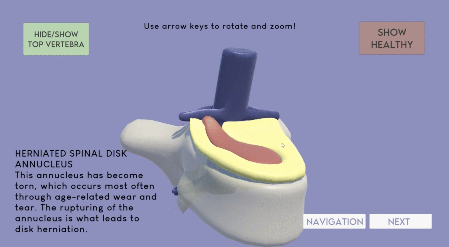
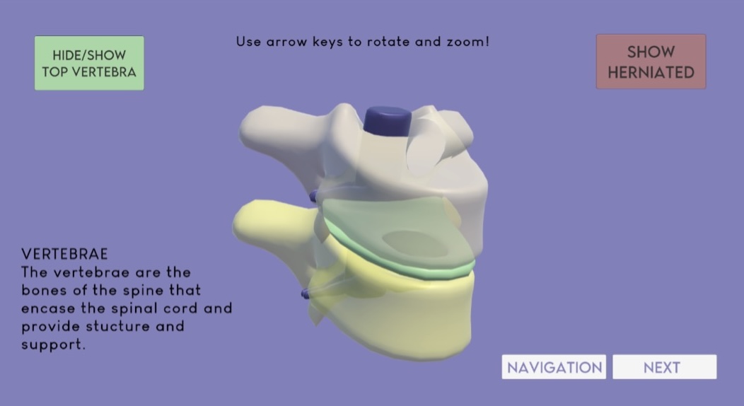
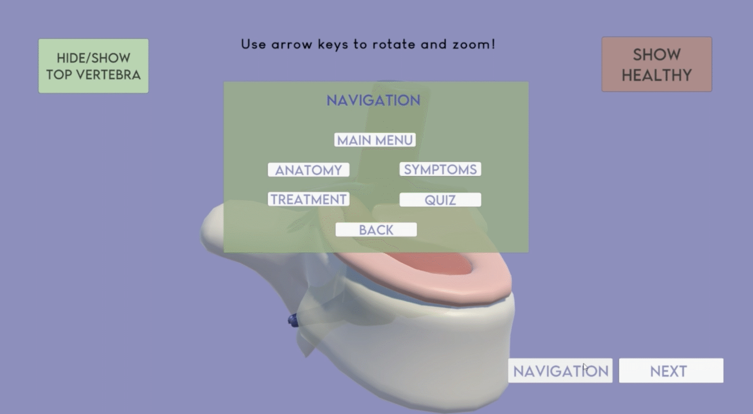
“Struck a Nerve” – Herniated disk explorer
This interactive application was developed in Unity 3D in collaboration with my classmates Ashna Konjeti and Chloe Levenson. Our goal was to create an engaging and simple learning tool for public education on herniated disks, a common spinal condition. My role in the project was to develop the disk anatomy explorer scene, where the user can zoom, rotate, and identify different anatomical areas of both healthy and herniated spinal disks. I was also responsible for creating a navigation menu that allows the user to navigate to any scene in the application, an important contribution to streamlining the user experience. Additions such as on-click colour changes and sounds enhanced the UI further. I really enjoyed the opportunity to enhance my skills in graphic design and UI for this project.
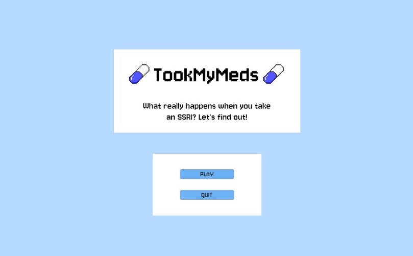
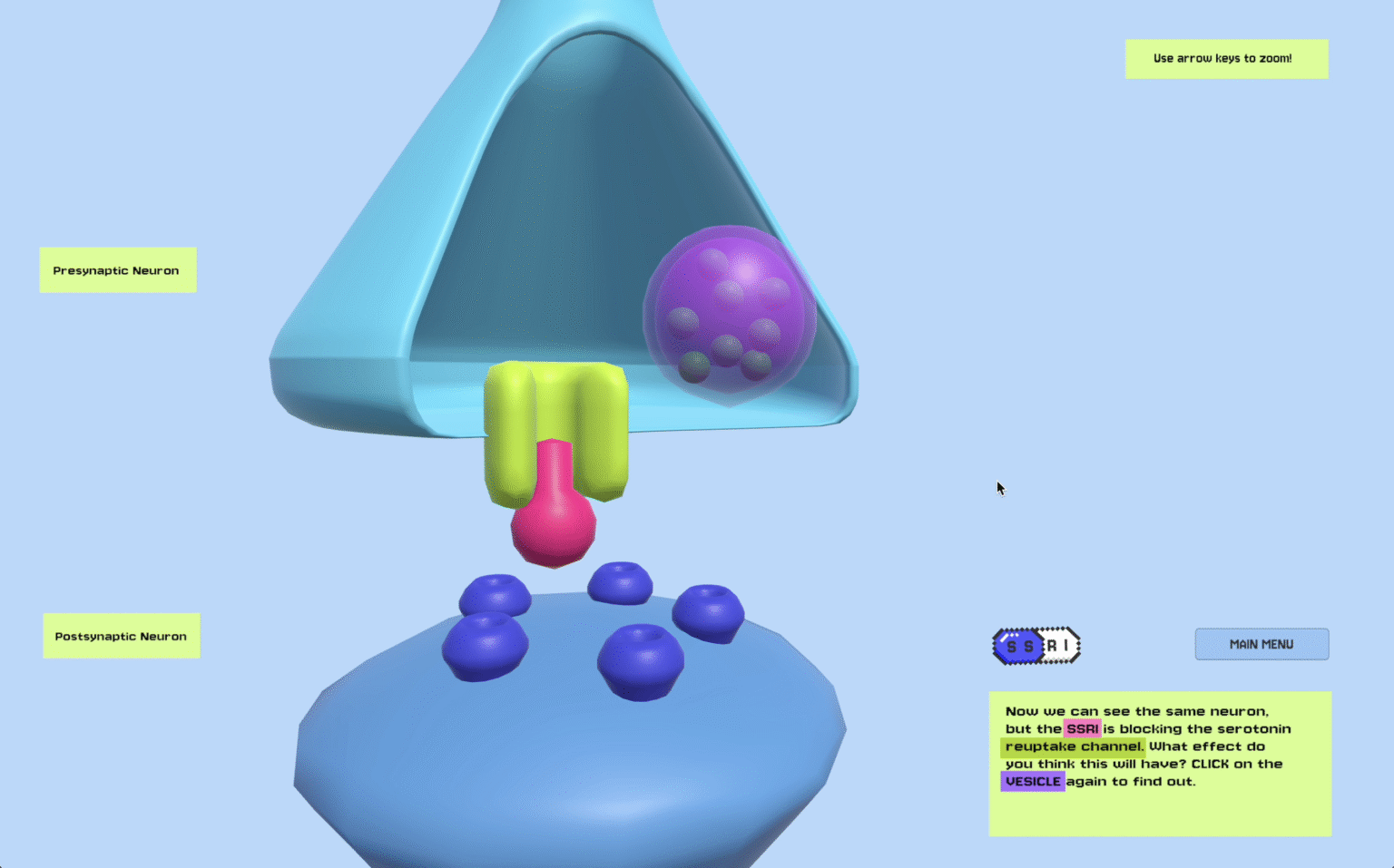
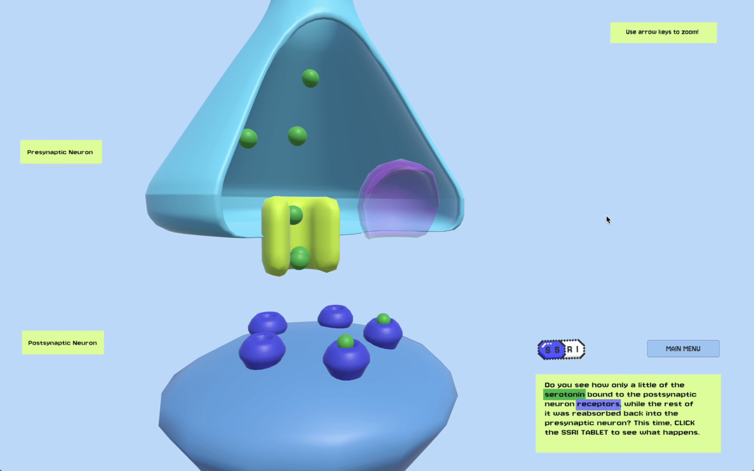
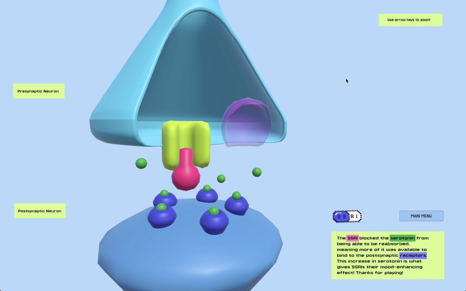
“TookMyMeds” – What really happens when you take an SSRI?
This interactive application was my first experience creating in Unity 3D. Inspired by my previous background in neuroscience, I opted to create a learning tool for simplifying and visualising the drug action of selective serotonin re-uptake inhibitors (SSRIs), a commonly prescribed type of medication used to treat depressive disorders. The application allows the user to release serotonin from the presynaptic neuron into the synaptic cleft, where it interacts with the postsynaptic neuron. The user is able to directly compare the action of serotonin in the synapse both without and with the presence of an SSRI.