MSc Medical Visualisation & Human Anatomy School of Innovation & Technology
Erin Armstrong
Hello, my name is Erin Armstrong, and I am a Biomedical Science with a Year in Industry graduate (BSc Hons) from the University of Sheffield. During my studies, I enjoyed gaining an understanding over a broad range of topics such as neuroscience, physiology, pharmacology and molecular biology. A pivotal experience for me was my placement year in which I joined the amazing team at Pebble Biotechnology Laboratories. I gained invaluable experience in the field of innovative medical technology, and got the opportunity to use my creativity and love or art to create some hand drawn diagrams and animations to help explain their work to patients and clients. This is what sparked my interest in combining my medical knowledge with visual technology, and led me to discover the master’s in Medical Visualisation and Human Anatomy.
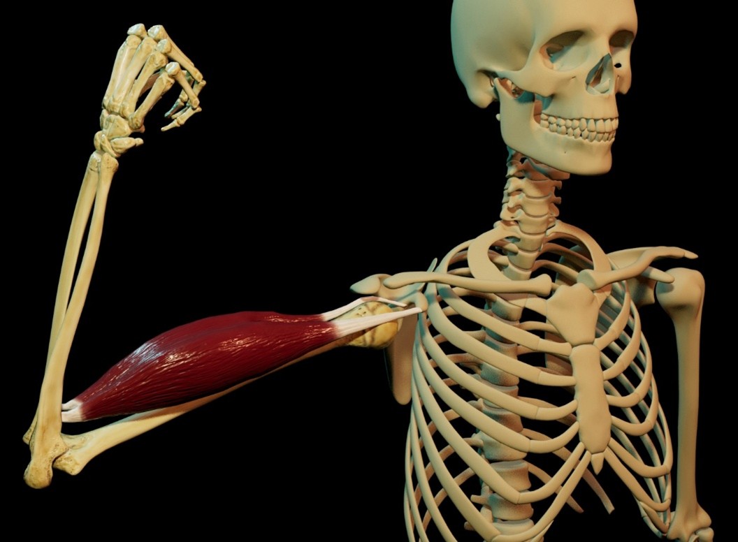
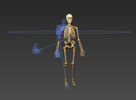
This year has been intense and challenging, but extremely rewarding. I have developed skills in a broad range of disciplines, including 3D modelling, animation, volumetric visualisation, interactive app development, and developing applications for extended reality. The MSc course has also helped to consolidate my anatomical knowledge through additional cadaveric dissections. For my thesis I collaborated with the University of Aberdeen to develop a mixed reality application aiming to help guide surgeons during free flap procedures. This was a great experience that allowed me to explore novel technology, interact with surgical professionals, and provide insight into a clear gap in medical research.
Interactive Visualisation
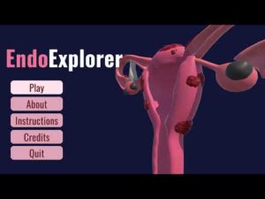
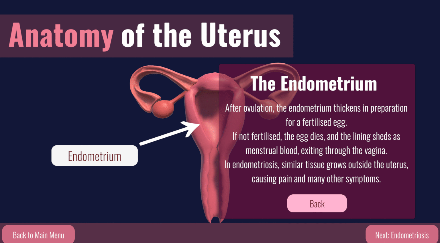
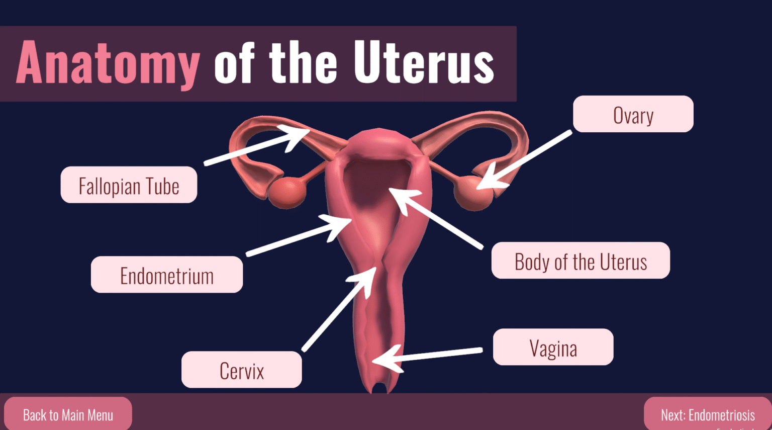
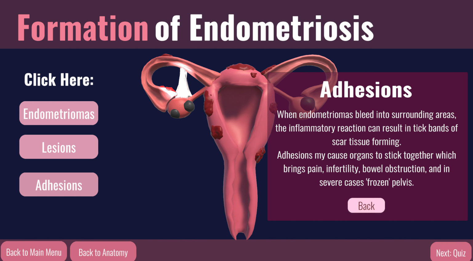
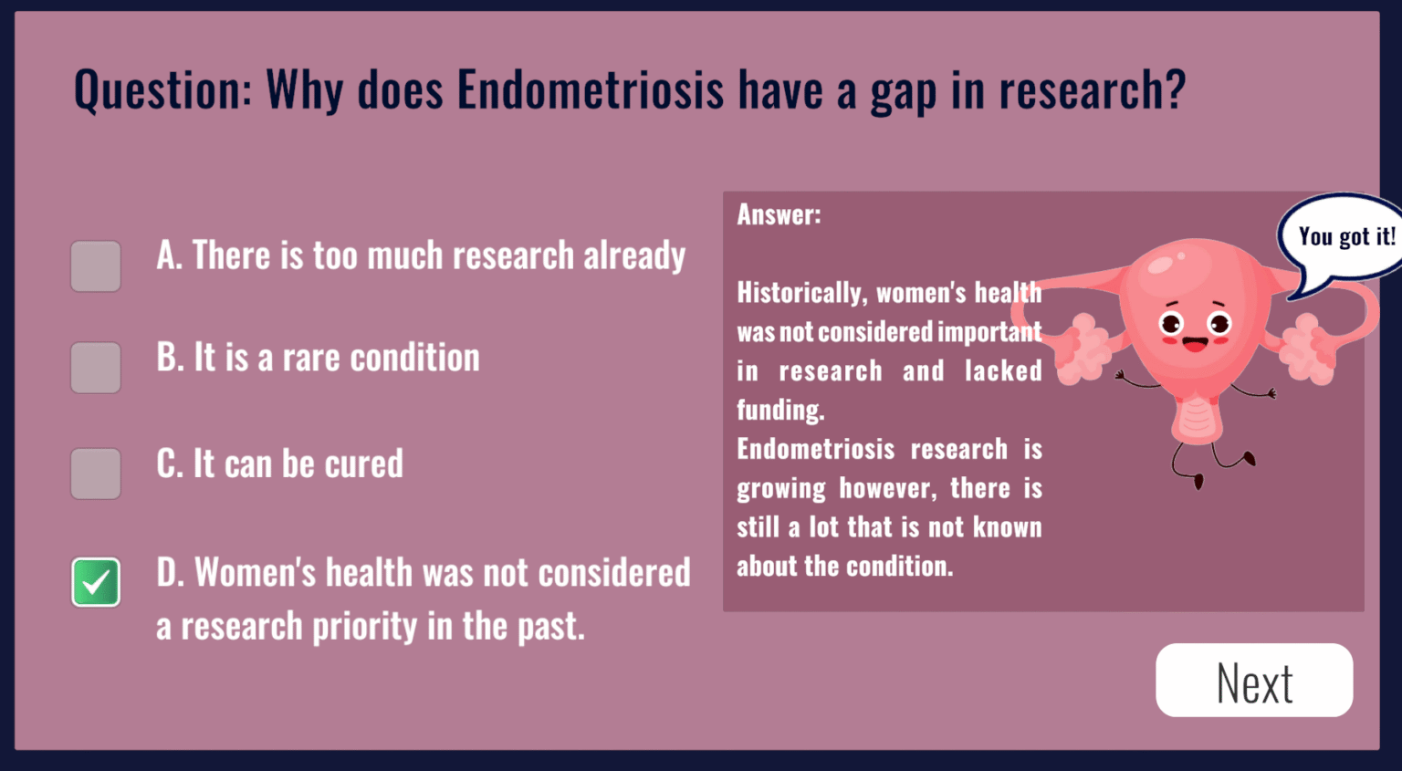
‘Endo Explorer’ was a collaborative project developed by myself along with two of my classmates Eilidh Stevenson and Rebecca Millar. The project aimed to develop an interesting and educational application focused on the topic of endometriosis. The application was developed in Unity and coded using C#.
Thesis Project
Precision in Focus: Evaluation of Oculus Meta Quest VR Headsets for Mixed Reality-Guided Free Flap Surgery.
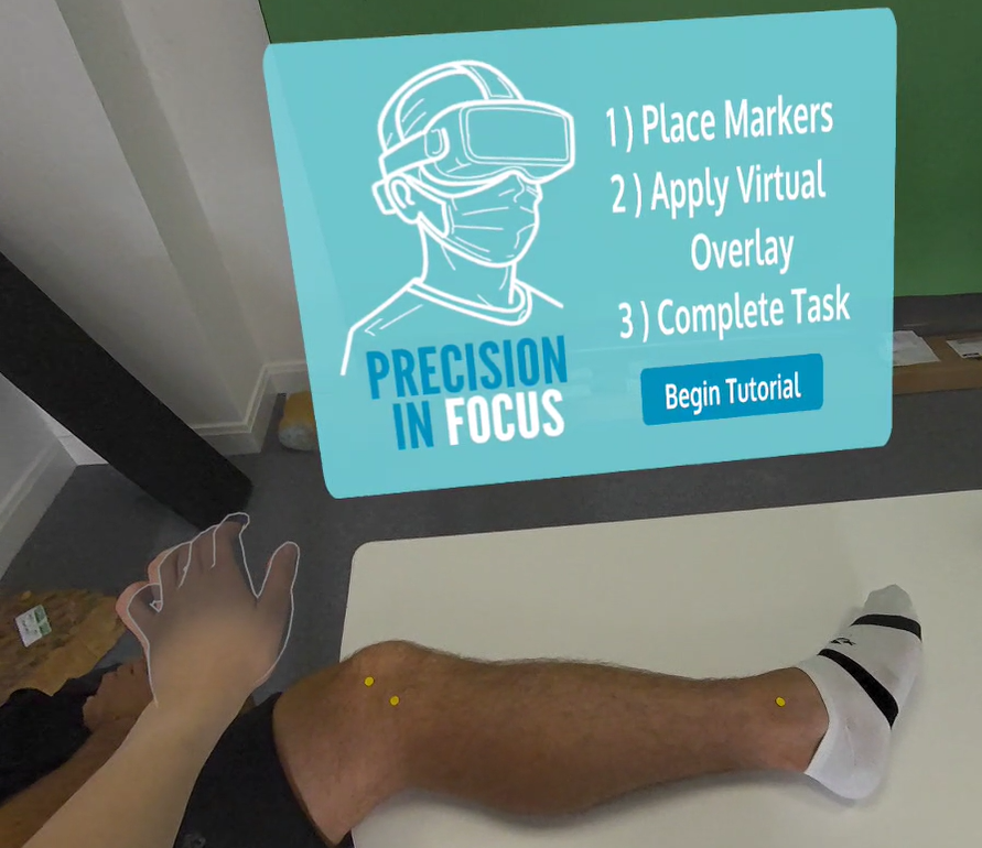
Augmented and mixed reality (MR) technologies enable patient-specific preoperative planning data to be overlaid directly onto the body intraoperatively, providing a real-time visual guide during surgical procedures. This is especially promising for high-precision reconstructive tasks such as free flap sectioning. This thesis project investigated the use of novel consumer-grade virtual reality (VR) headsets with MR capabilities, the Oculus Meta Quest series, which are largely unexplored in surgical overlay contexts. These headsets have features that could potentially overcome the challenges seen with other widely explored headsets; such as limited accuracy and visual perception issues, which have hindered clinical adoption to date.
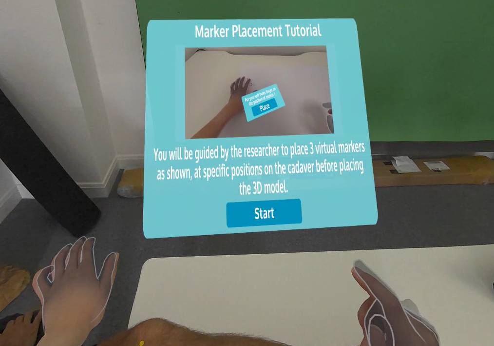
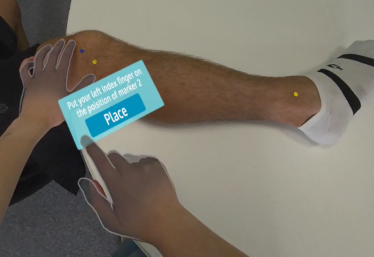
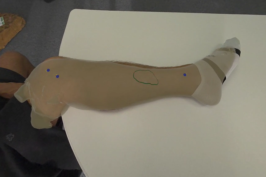
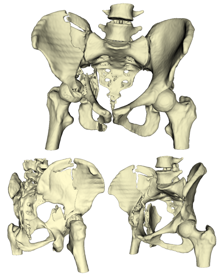
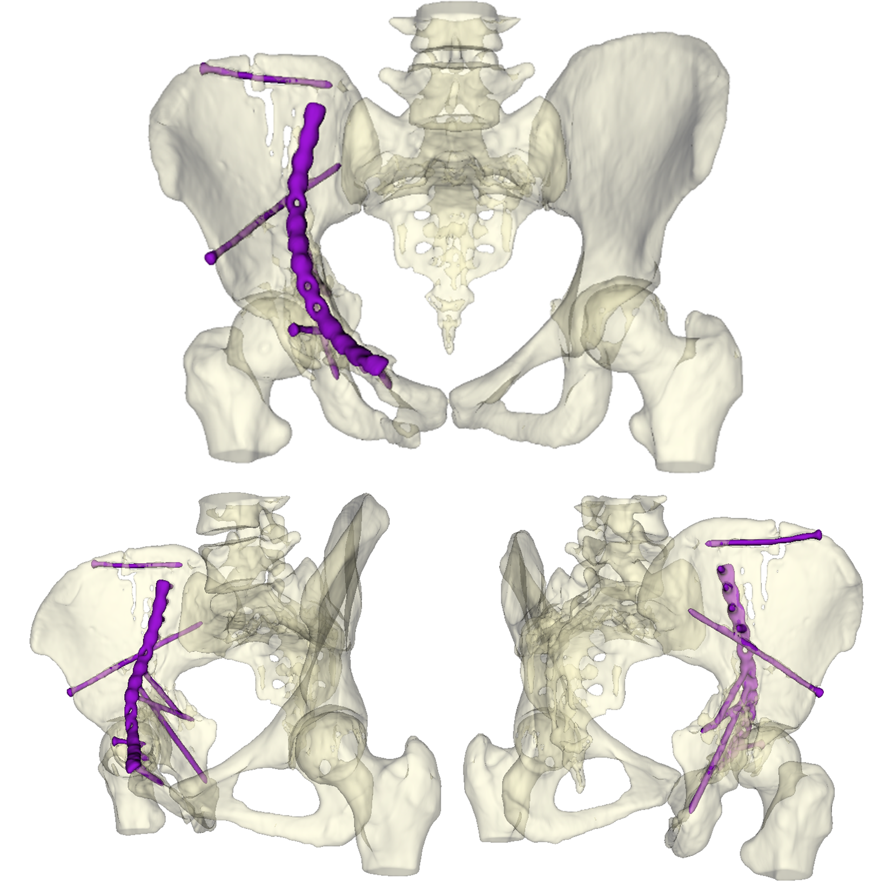
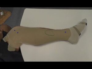
The project involved the exploration of novel tracking and registration techniques, to overlay a virtual 3D model of a planned flap margin onto a cadaveric leg. Hand-tracking enabled the placement of three virtual markers at predefined positions on the leg, these markers were then used to calculate the correct position and rotation of the 3D model and spatially anchor it in the mixed reality environment. This system was employed on the Oculus Meta Quest 3 and evaluated in a pilot feasibility study using a group of surgeons from the Aberdeen Royal Infirmary.
This project was conducted in collaboration with the University of Aberdeen, and I would like to thank everyone who contributed to or took part in this study. I would also like to express my gratitude to my supervisors, Dr Matthieu Poyade, Prof. Flora Groening, Dr Eilidh Ferguson, Dr Jenny Clancy, and Dr Jenny Gregory for their continuous support and guidance throughout the project.
Video Walkthrough of the Application
This 3D model was generated from CTA scan data of a cadaveric leg, and was intended to be overlaid onto that specific specimen. However, due to the sensitive nature of using a cadaver in this study, this video walkthrough of the application has been demonstrated on a healthy volunteer's leg to help visualise the concept.