MSc Medical Visualisation & Human Anatomy School of Innovation & Technology
Elizabeth Anne Dunlap
Blending my work as a figurative artist, self-published author, and clinical bodyworker, I have spent more than twenty years practising bodywork and teaching functional movement, refining ways to communicate essential principles to my clients more effectively.
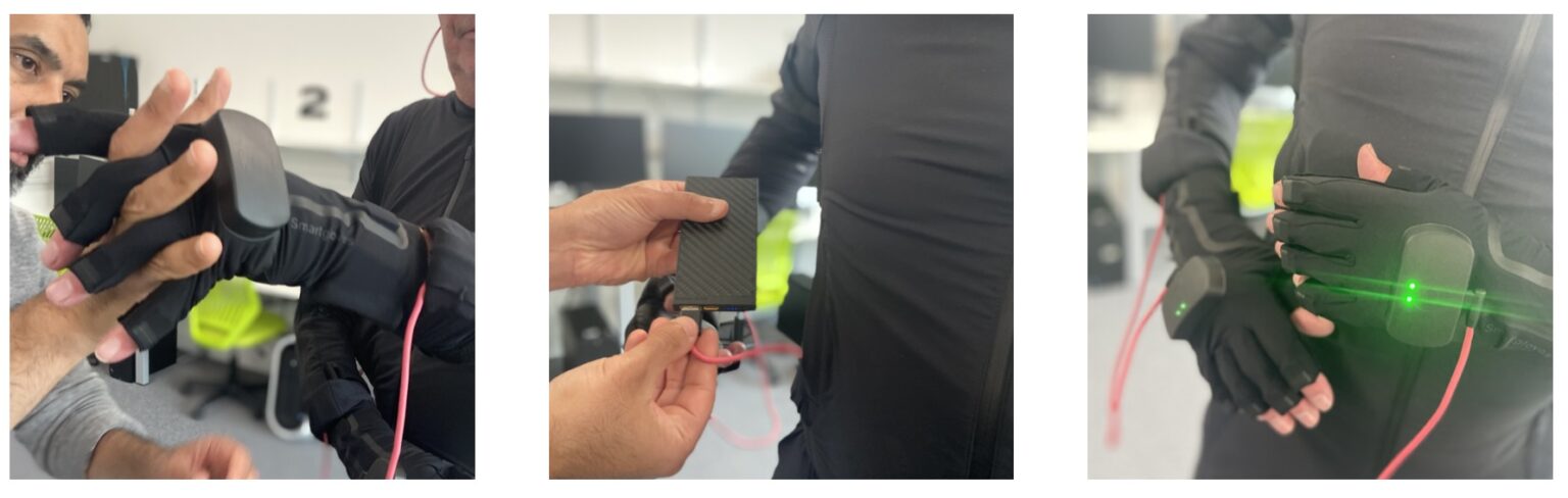
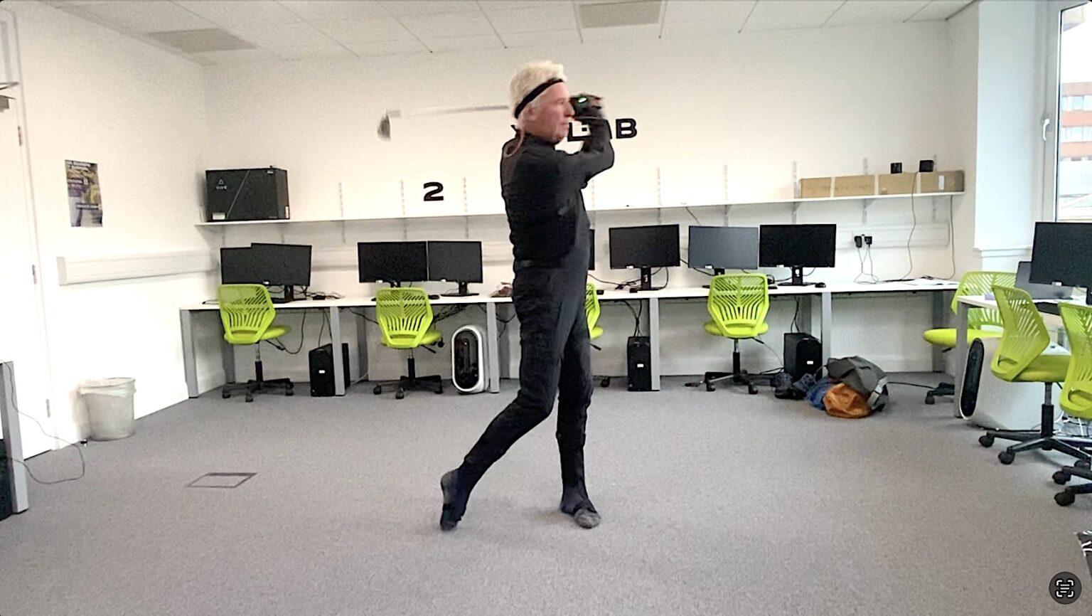
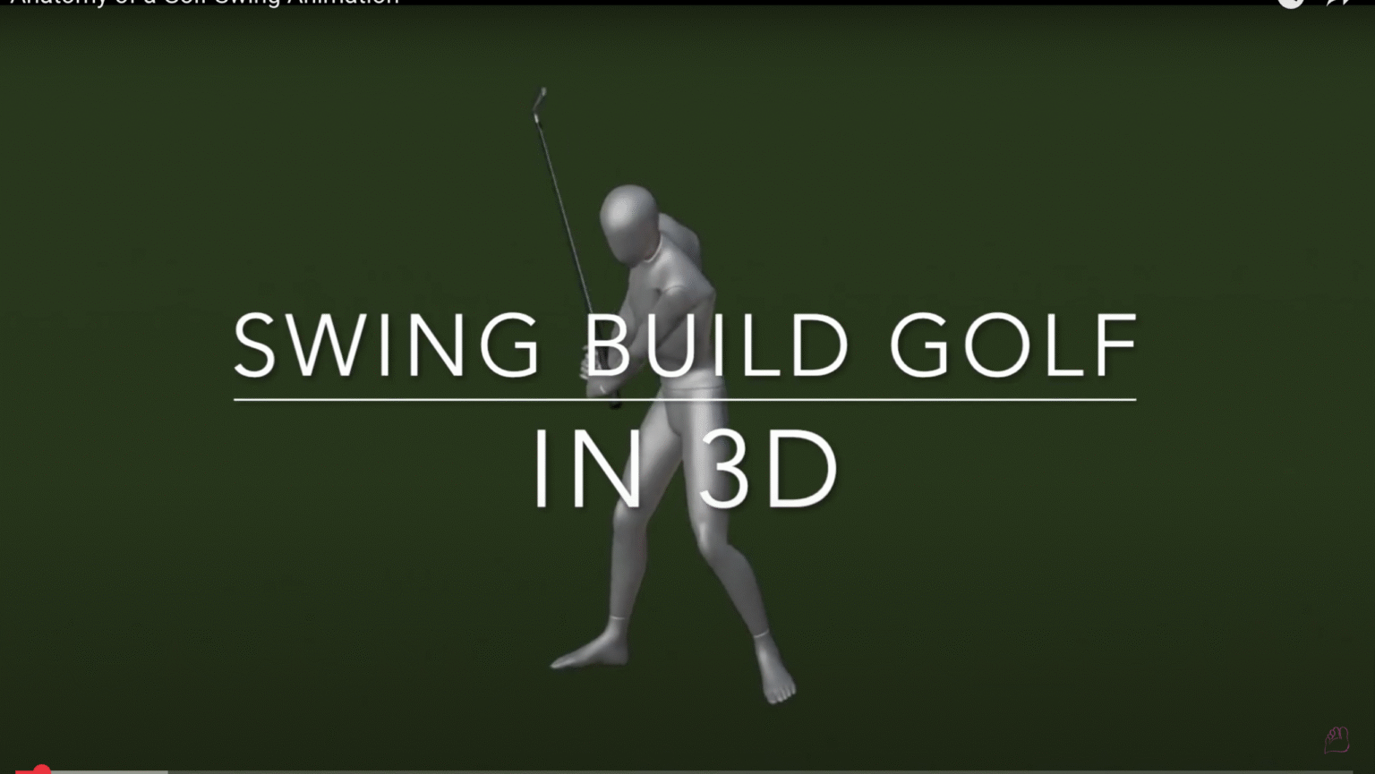
Crafting my master’s study, Medical Visualisation of the Golf Swing, in collaboration with Swing Build Golf, I created an anatomically accurate 3D animation that reveals skeletal and muscular movement during foundational swing exercises. Built from motion capture data, the project distils complex swing mechanics into a clear, repeatable learning tool for golfers and coaches.
Expanding my focus, I plan to integrate 3D modelling and animation into future movement education projects, using medical visualisation and motion capture to connect felt experience with anatomical understanding.
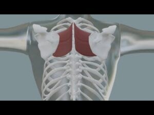
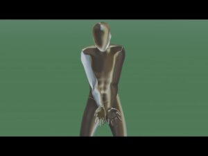
Anatomy of a Golf Swing
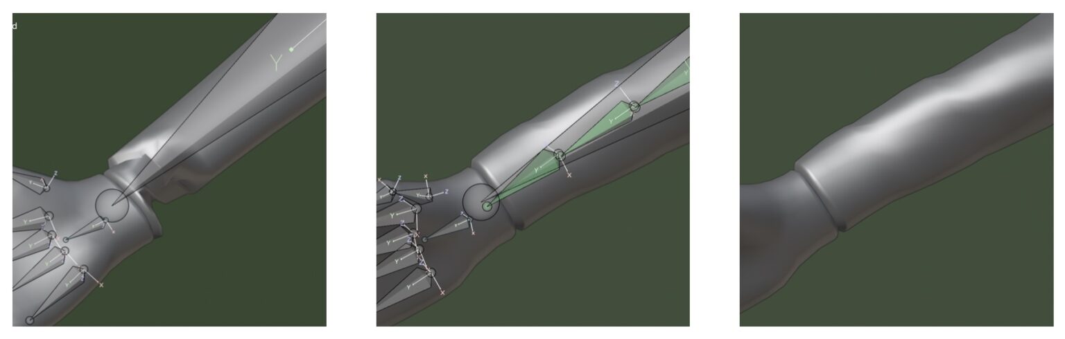
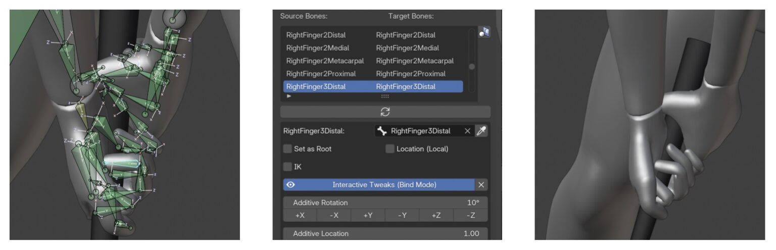
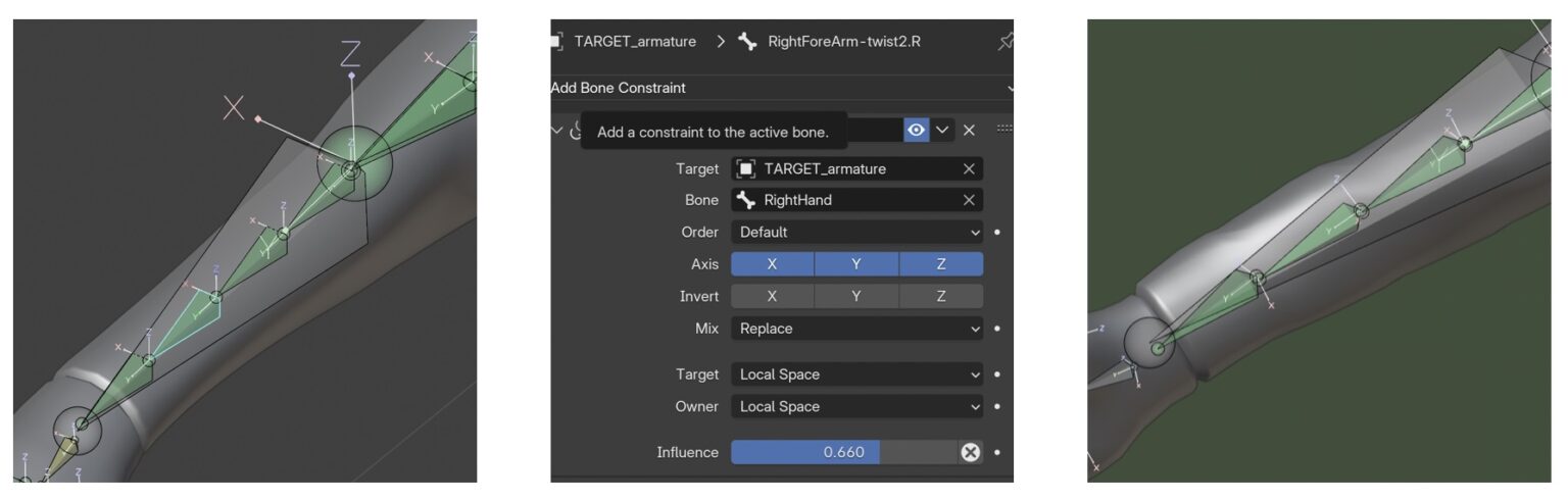
This 3D anatomical animation visualises three foundational exercises from the Swing Build Golf method. Created in Blender 4.4.1, it incorporates motion capture data recorded at the Glasgow School of Art XR Lab, retargeted to a 3D character model. The rigging of the target model was manually adjusted to improve arm deformation, and additive animation refined the movement of the hands. The skin was rendered with a transparent material to reveal muscle and bone movement, and with a silver metallic material to show the kinesiology of the body mechanics. The final project offers golfers and coaches a precise, repeatable visual reference for understanding coordinated swing movement
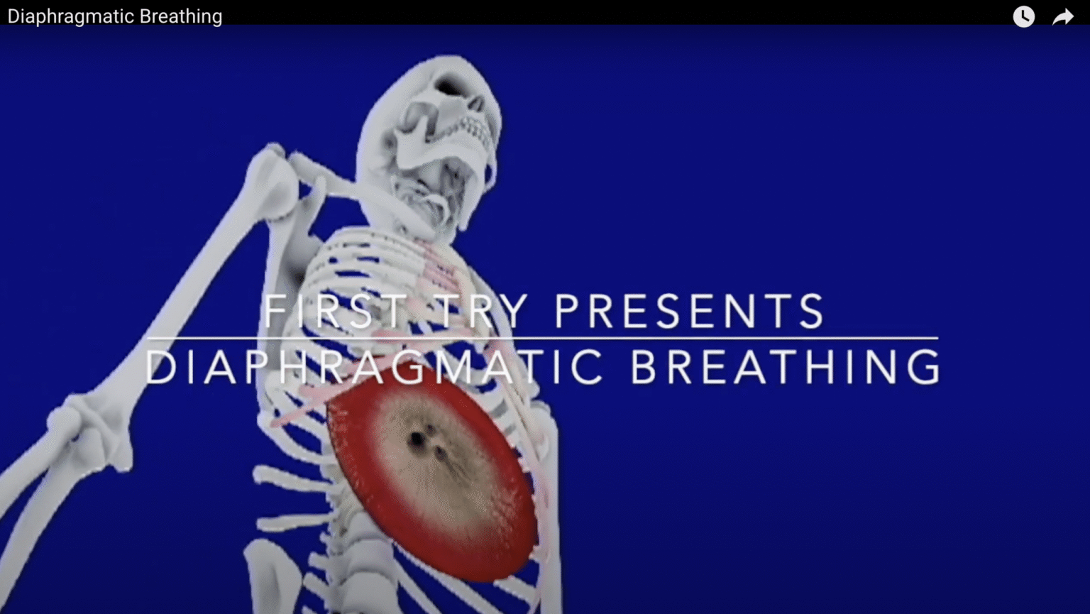
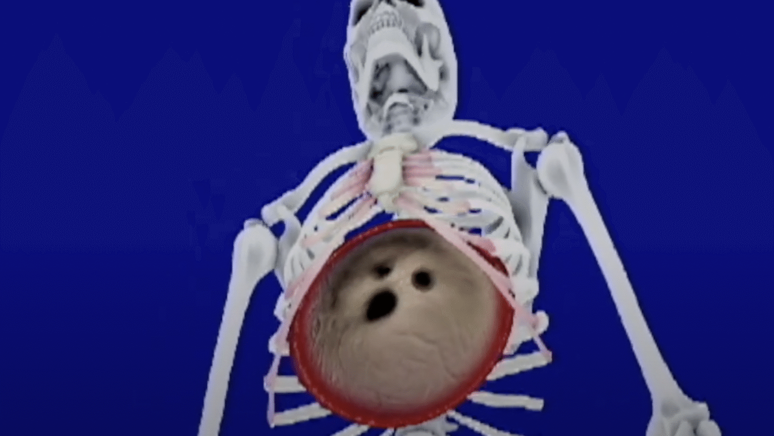
Diaphragmatic Breathing
This first-semester 3D modeling and animation project depicts diaphragmatic breathing, showing the contraction and relaxation of the diaphragm in relation to the rib cage and thoracic vertebrae. The diaphragm was sculpted in ZBrush, while the rib cage and vertebrae were retopologised and animated in 3D Studio Max. Retopology was performed manually in 3D Studio Max over several weeks — a time-intensive approach that, in the hands of this artist, led to rendering issues and a low-resolution final output. UV unwrapping also proved challenging and was ultimately completed in ZBrush.
These outcomes reflect the artist’s early-stage workflow decisions rather than any limitation of the software. For students intending to work extensively in 3D Studio Max, a PC is strongly recommended over a Mac for compatibility and performance. With more experience, Blender has since become my preferred tool for its integrated, Mac-friendly environment, reducing the need to move between programs; in my opinion, it may eventually replace 3D Studio Max and ZBrush in many workflows. That said, I also believe the renders achievable with 3D Studio Max and ZBrush remain superior in quality. The final animation presents the functional role of the diaphragm during respiration.
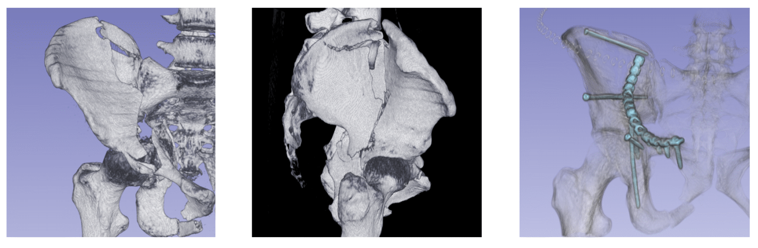
Volumetric Visualisation
This visualisation contains medical imagery of a pelvis before and after surgery, including fractures, surgical hardware, and sutures. While presented in a softened, sketched style, the images may still be distressing to some viewers. Viewer discretion is advised.
Converting medical scan data into 3D models using 3D Slicer via Direct Volume Rendering, I visualised a human pelvis before and after reconstructive surgery. Black-and-white images with a sketched look help reduce the visual impact of the injuries for the sake of the patient. I chose to keep visible the area where sutures are indicated, giving the patient a clearer understanding of the surgical work performed. The pre-surgery model reveals a severe fracture on the right side, with the pelvic ring broken and bone fragments displaced. The post-surgery model, rendered with semi-transparent bone, draws attention to the plates, screws, and rods used to reconnect and stabilise the pelvis while maintaining a softened presentation.
Medical Visualisation of a Golf Swing – Developing and Evaluating a 3D Anatomical Animation
Golf attracts more than 100 million participants worldwide. There is clear demand for innovative and accessible ways to support skill development. Two aspects make this project distinctive. First, it shows the skeletal and muscular system in motion, linking swing mechanics to anatomy. Second, it embeds narration from an established coach, combining visualisation with lived teaching practice. The final animation runs four minutes twenty-three seconds, exported in 1920 x 1080 format, and deployed privately on YouTube with closed captions for accessibility.
Evaluation used a mixed-methods approach.
Qualitative feedback: Participants praised sequencing, rhythm, and timing cues, and described the animation as clear and usable. They also suggested slower pacing, clearer tempo guidance, and visual consistency between club and handedness.
Quantitative results: Simulator testing focused on carry distance and club head speed. Across all five swings, no significant change was detected. But when best-of-three swings were analysed, three of four t-tests indicated statistically significant improvement, with reduced variability in performance.
The project resulted in a modular, replayable digital learning tool that visually translates the Swing Build method into a portable format. It established proof of concept for using medical visualisation in golf instruction.