MSc Medical Visualisation & Human Anatomy School of Innovation & Technology
Eilidh Stevenson

Hello! I’m Eilidh, an Anatomy BSc (Hons) graduate from the University of Glasgow, with a particular interest in medical imaging, the cardiovascular system and improving healthcare through visualisation. After graduating, I worked within the Anatomy Facility as a demonstrator, where I attended a guest talk by Daniel Crawford, the founder of Axial3D– a pioneering company in the field of medical visualisation. This initiated my interest in the field, and as someone who had always loved both art and science, MSc Medical Visualisation & Human Anatomy felt like the perfect combination!
This year has been an exciting and challenging opportunity to develop my anatomy knowledge and learn a range of technical visualisation skills, including: Volumetric Visualisation, Interactive Visualisation, Immersive Technologies/Human Computer Interaction and 3D Modelling. For my thesis project, I had the privilege of working with the NHS Nuclear Cardiology Department at the Glasgow Royal Infirmary to improve patient communication, with the aim of improving understanding and reducing anxiety for patients. This involved creating an interactive resource for patients attending nuclear cardiology for myocardial perfusion imaging.
Throughout the course, I particularly enjoyed learning volumetric visualisation techniques to visualise anatomy from imaging datasets. I am excited to apply my knowledge as I begin working at Axial3D as an Associate Production Engineer– using this skillset for patient-specific care and surgical planning.
I would like to thank Dr Matthieu Poyade, Kerri Thornton and Danny Buksh at GSA for making a very steep learning curve an exciting and enjoyable experience. I would also like to thank the wonderful staff at the Anatomy Facility, without your encouragement and support since 2020, I would not have pursued MedVis.
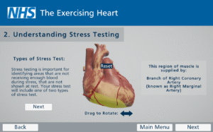
Thesis Project: The Exercising Heart
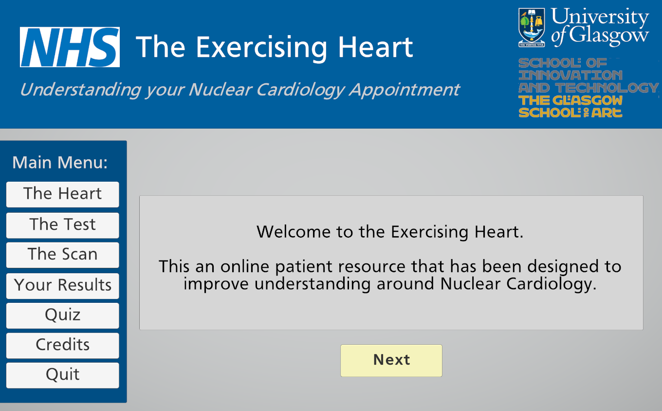
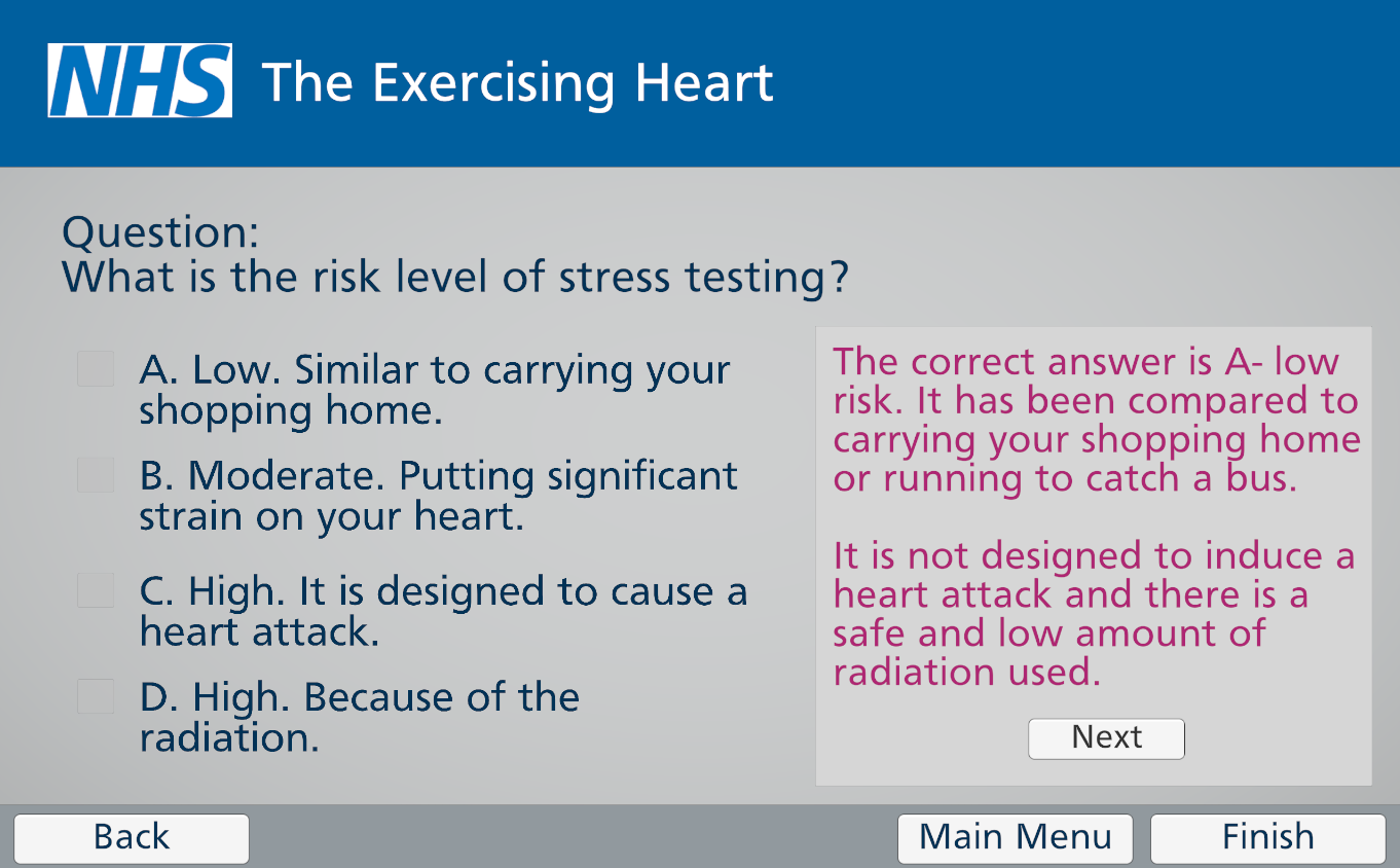
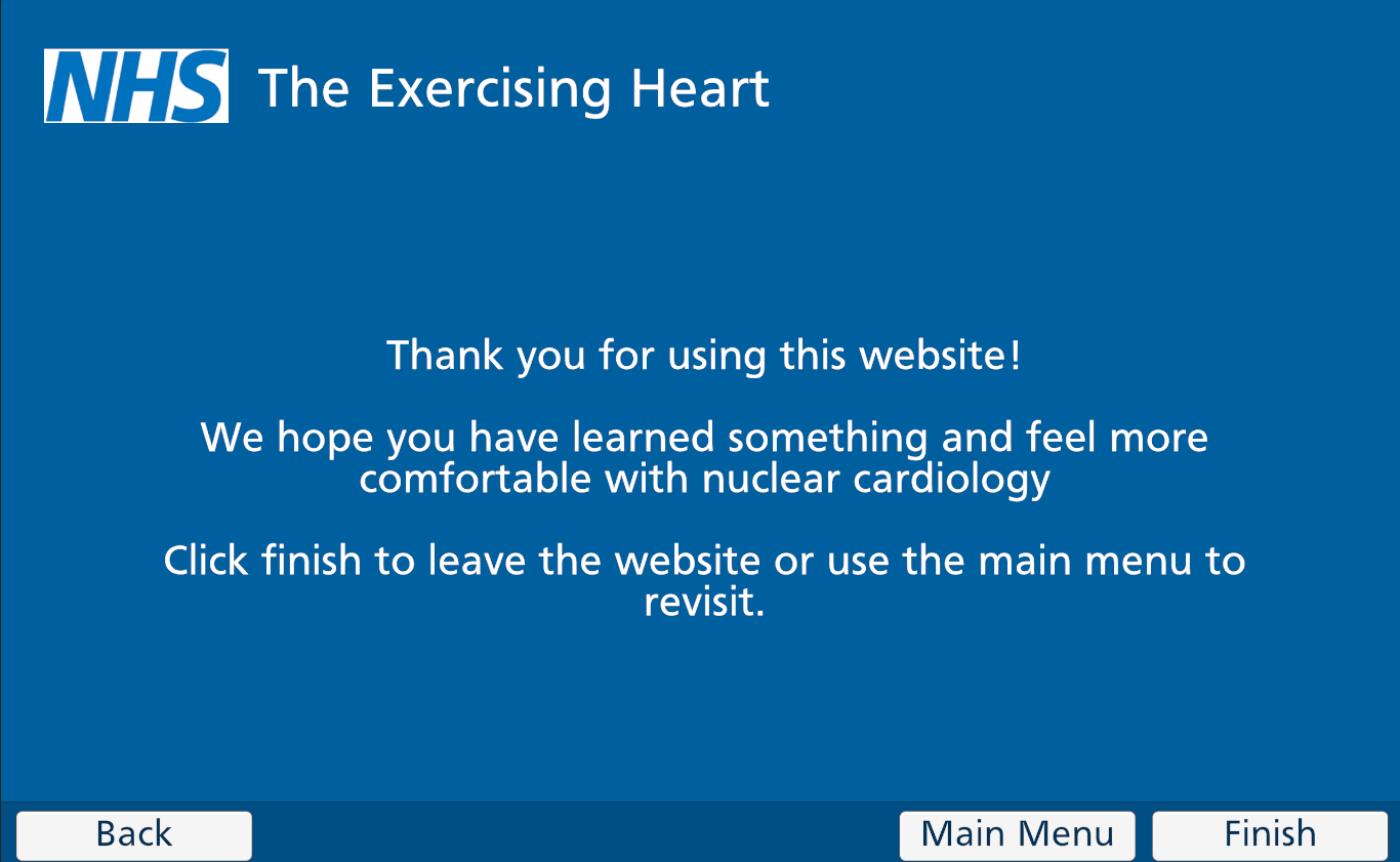
The Exercising Heart: An Interactive Web-Based Application for NHS Nuclear Cardiology Patients
This project was completed in partnership with the NHS Nuclear Cardiology Department at the Glasgow Royal Infirmary to improve patient communication. The rationale for this project was that patients often arrive at nuclear cardiology appointments feeling anxious or stressed due to misinformation and misconceptions around nuclear medicine, with NHS resources for nuclear cardiology being limited.
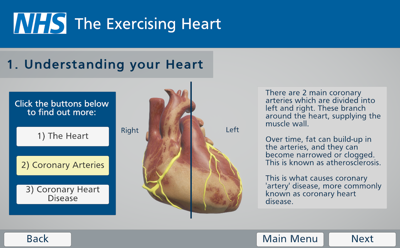
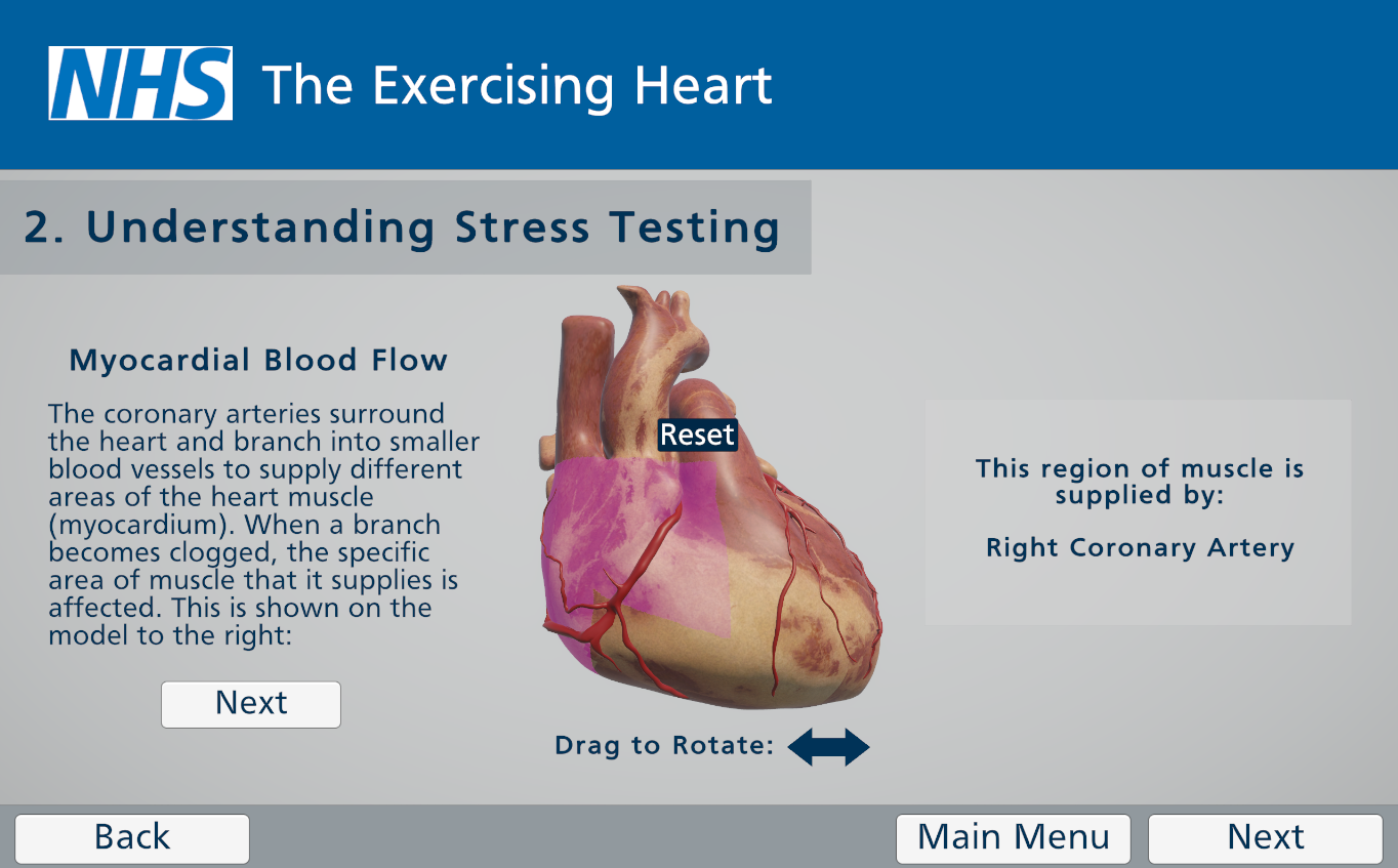
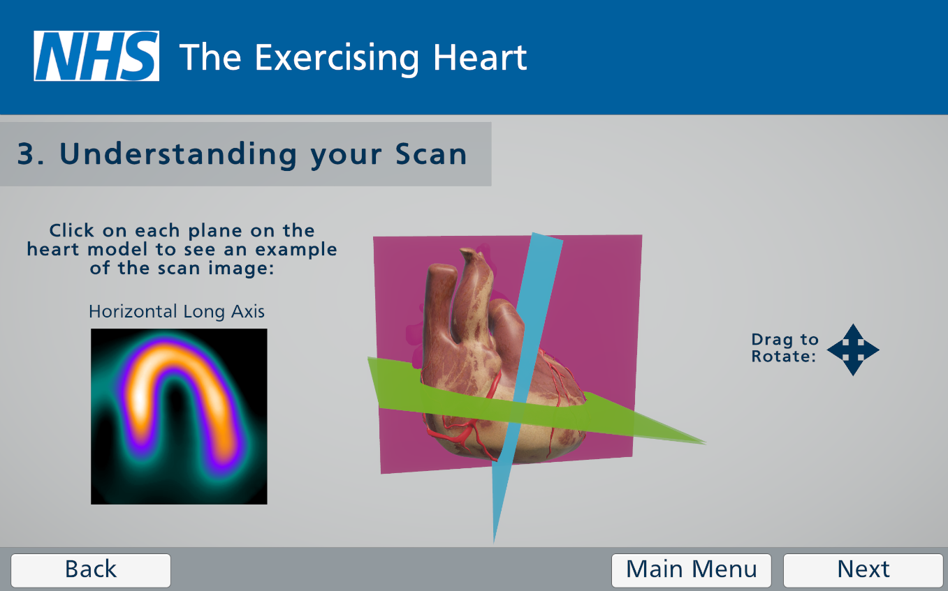
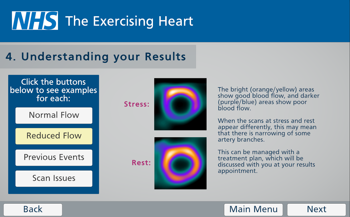
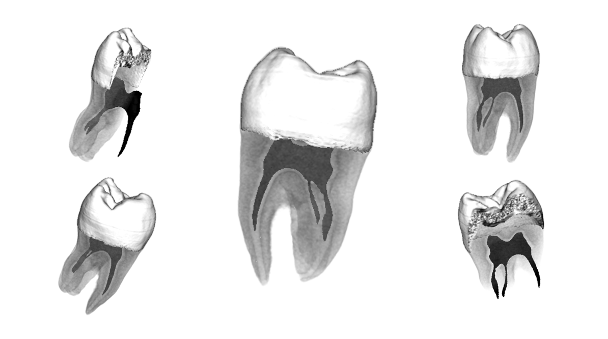
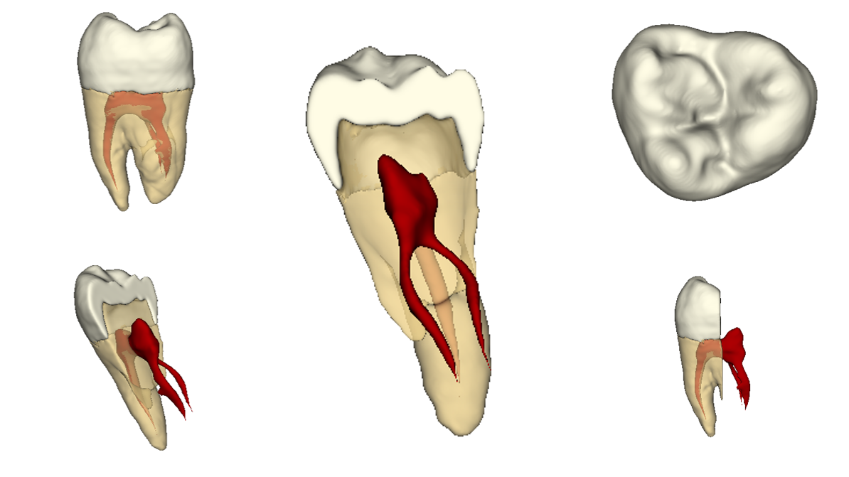
This gap in patient communication was addressed through the development and implementation of an interactive web-based application in a patient population. Complex nuclear cardiology procedures including stress testing and myocardial perfusion imaging were explained alongside an anatomical model of the heart created for the project. Various interactive elements were also developed to improve user engagement. Overall, the application was found to improve understanding of nuclear cardiology and reduce patient anxiety.
I would like to thank the wonderful supervision team for this project: Dr Matthieu Poyade, Dr Emma Bailey, Dr Andrew Carradus, Dr Kirsty Jones and Ms Barbara Kerr, for their encouragement and guidance throughout. I would also like to thank the staff within Nuclear Cardiology at the Glasgow Royal Infirmary for welcoming me into the department and accommodating testing. Finally, a huge thank you to the patients attending nuclear cardiology for their participation and feedback.
3D Modelling and Animation
This module required the use of 3D modelling and animation software including 3Ds Max and ZBrush, alongside visual effects software such as Adobe After Effects and Media Encoder. This also required the use of other Adobe Suite products such as Photoshop. Coming from a scientific background, I found this portion of the course daunting however, the final outcomes were really rewarding!
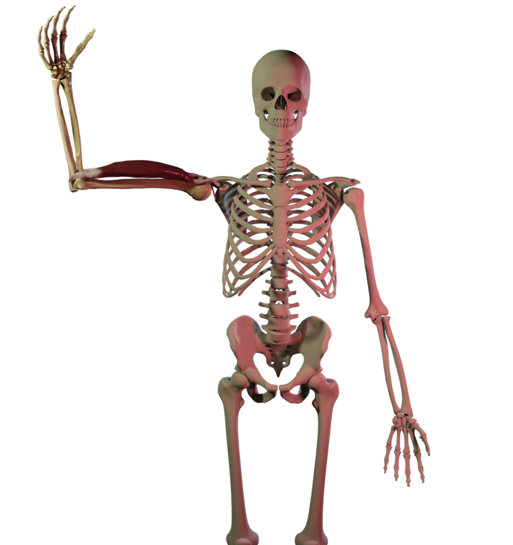
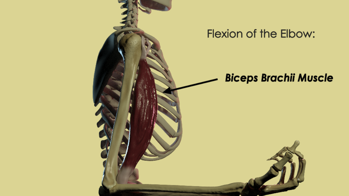
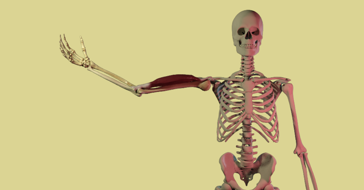
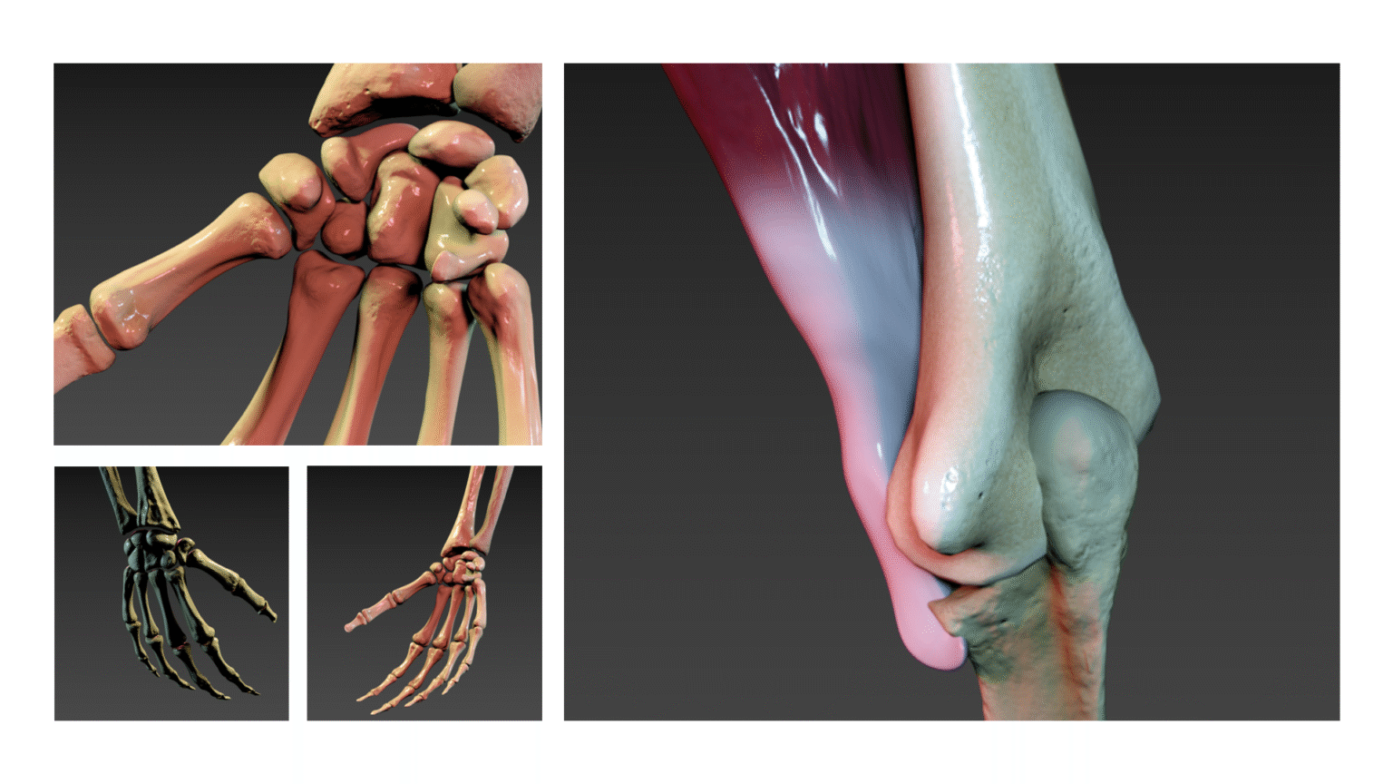
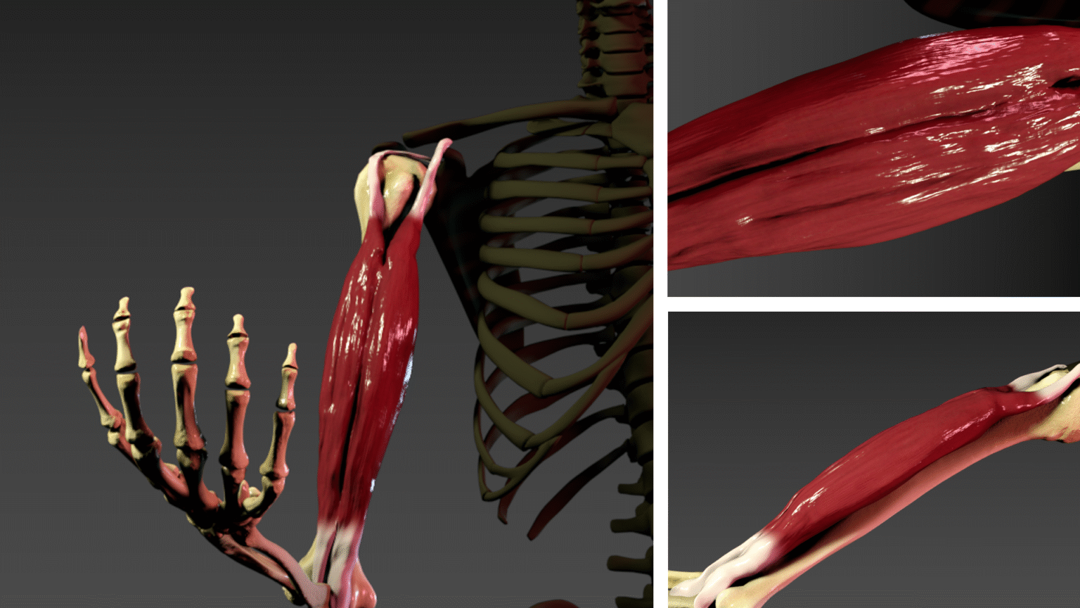
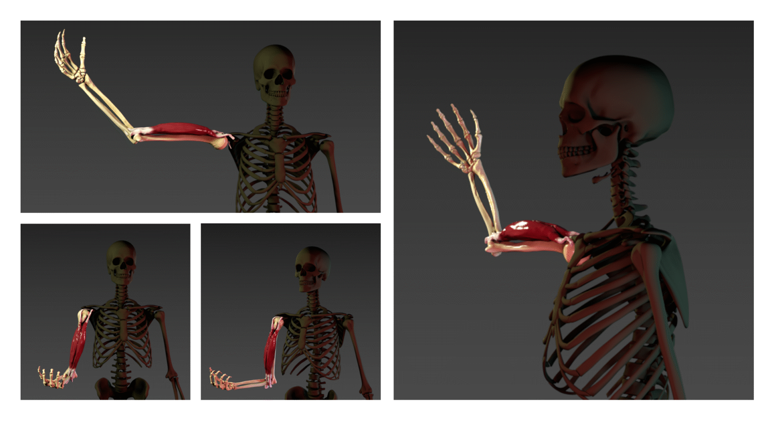
My final project was titled ‘Anatomy of the Upper Limb: A Short Animation’. This involved the re-topology, sculpting, texturing and poly-painting the bones of the upper limb, before creating a muscle of choice. The biceps brachii muscle was modelled and adjusted to the skeleton, before animating the whole upper limb. After rendering, this was imported to Adobe After Effects and Media Encoder for visual effects.
Interactive Visualisation
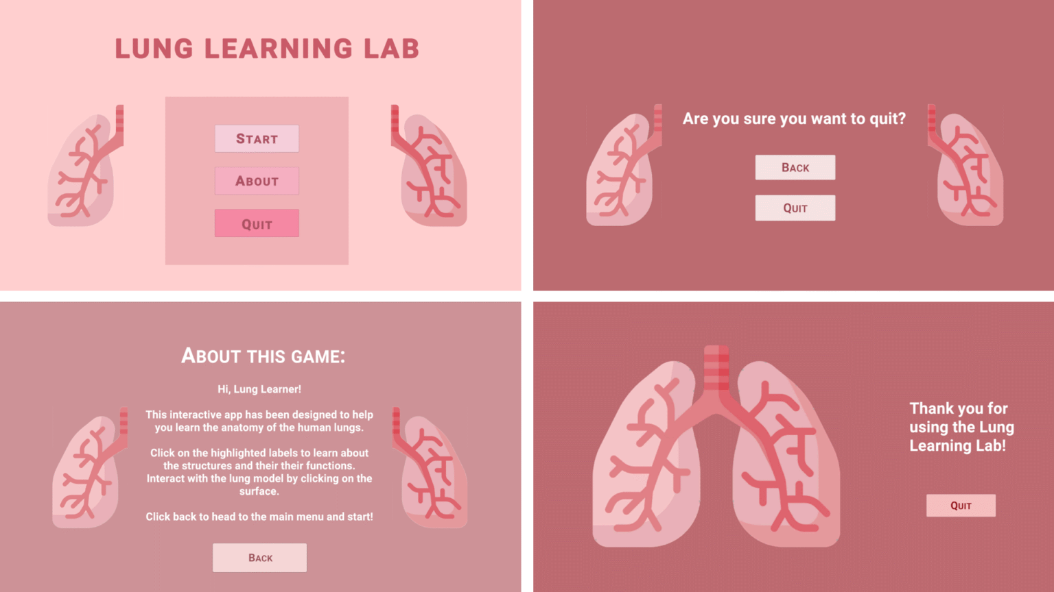
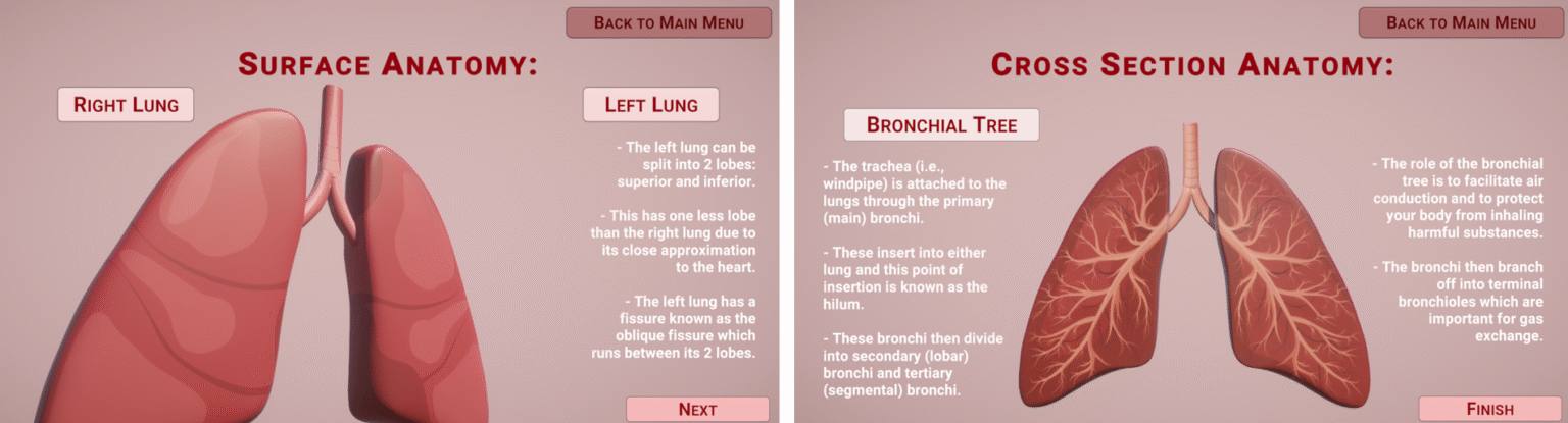
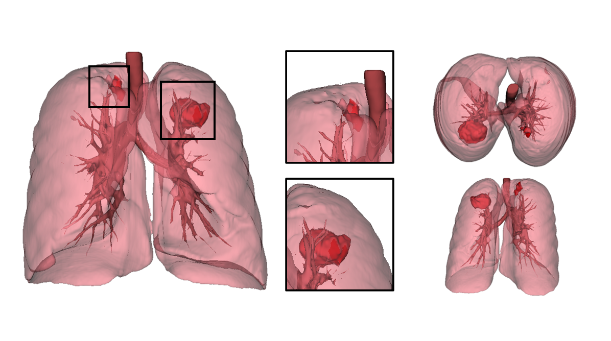
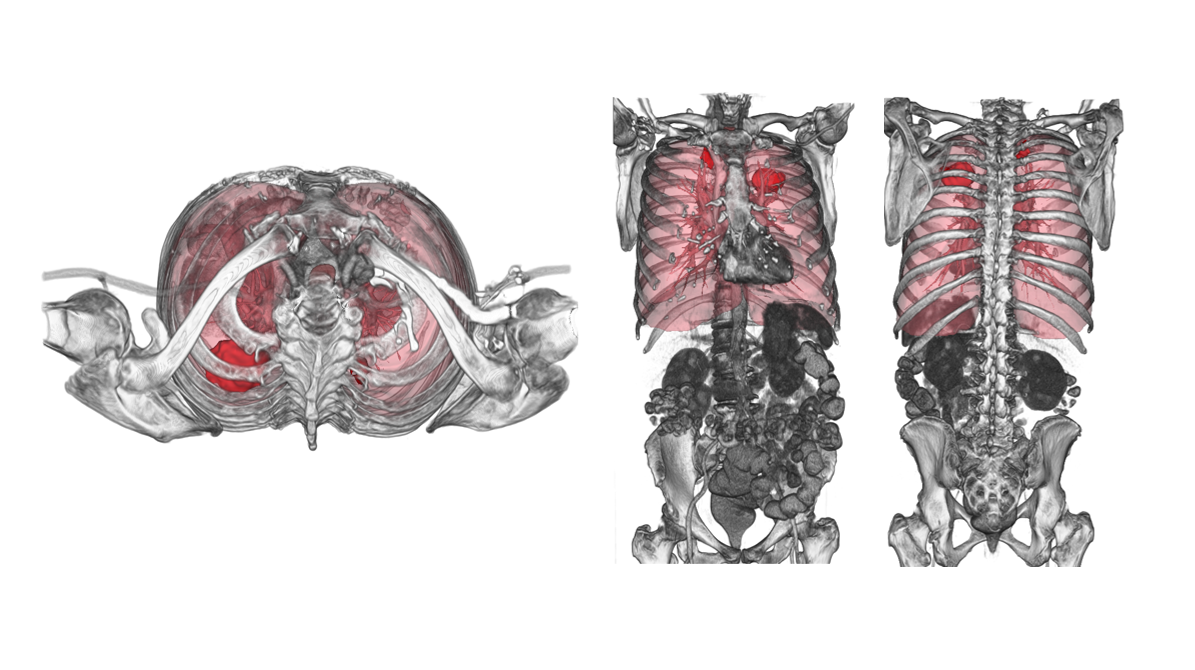
This module focused on developing interactive applications using Unity Engine, with coding using C#. The first development, ‘Lung Learning Lab’, was created for the Diorama project and was a short application introducing basic anatomy about the lungs.
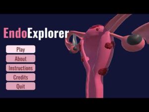
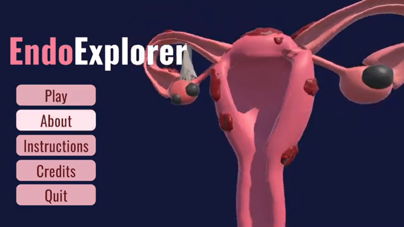
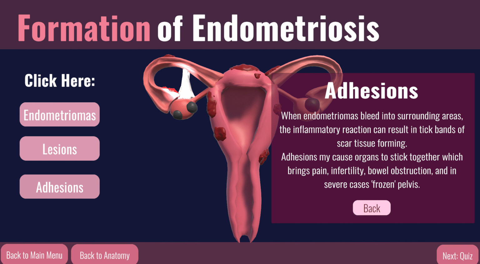
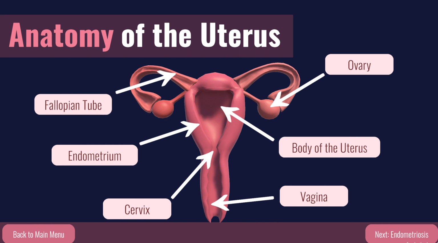
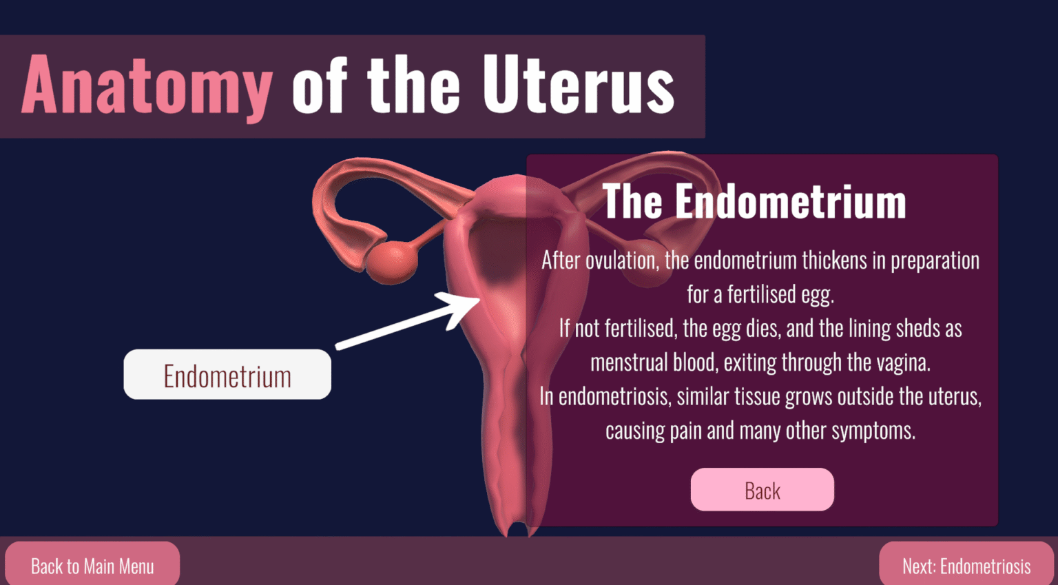
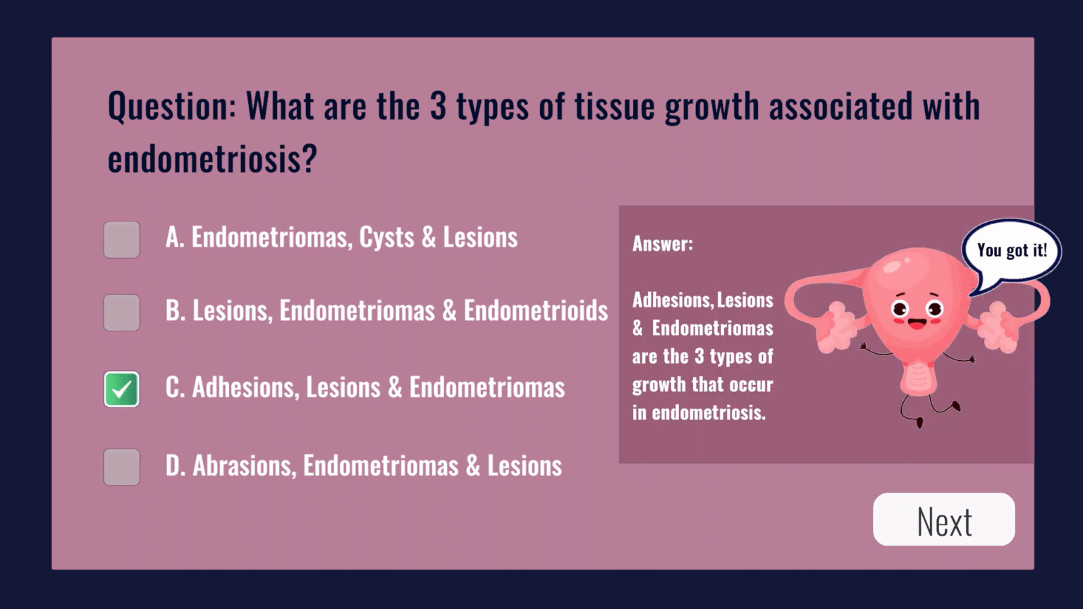
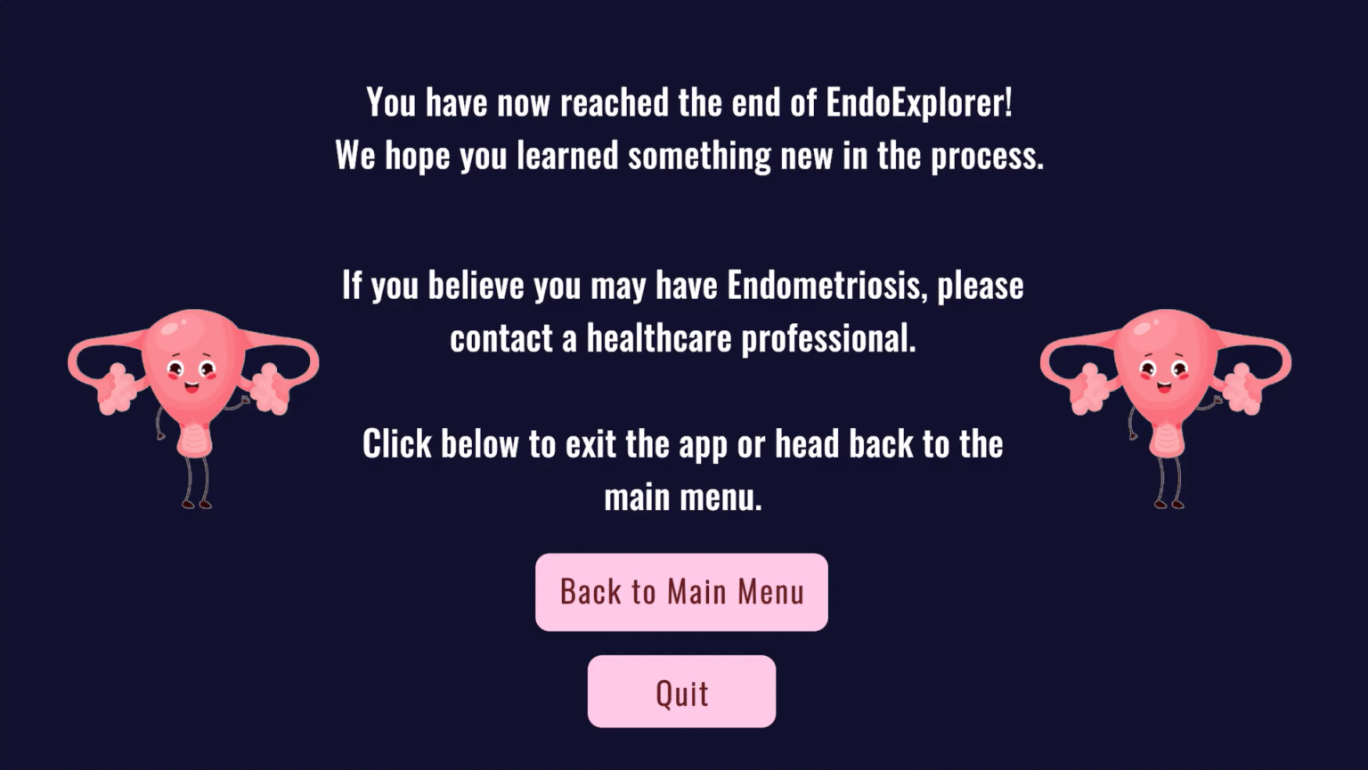
The final project was created in collaboration with my amazing and talented colleagues Erin Armstrong and Rebecca Millar, titled ‘EndoExplorer’. GitHub was used for version control, allowing tracked changes, important when developing in the industry.