Aqsa Akhtar
(She/Her)
Hello! My name is Aqsa Akhtar, an MSc Medical Visualisation and Human Anatomy student. My background is in (BSc) Immersive Systems Design – Games and Virtual Reality previously completed at The Glasgow School of Art.
I have gained many new skills throughout this course and had the amazing opportunity to collaborate with the Lancashire Teaching Hospitals NHS Foundation Trust (LTHTR) for my research project.
Check out some of the work I completed during this course below!
VR MRI Application – MSc Medvis Research Project
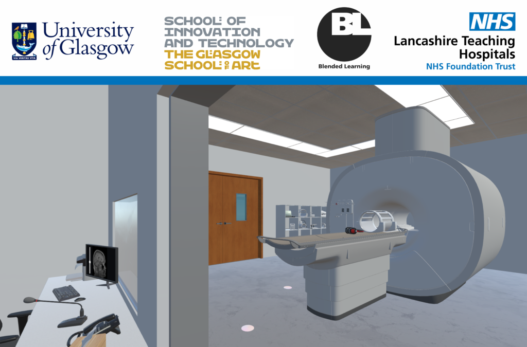
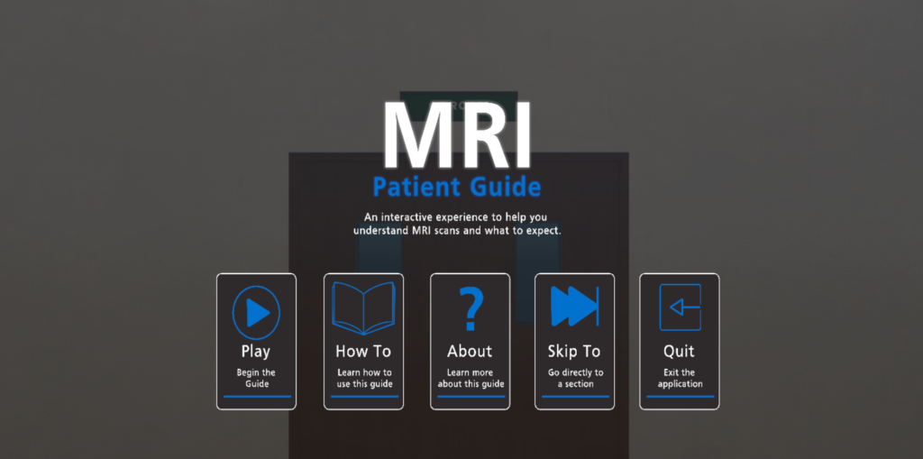
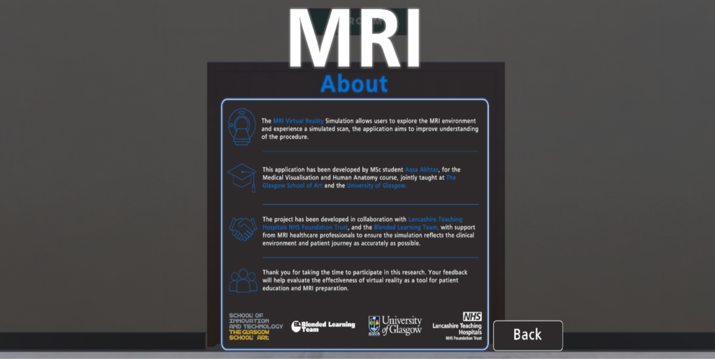
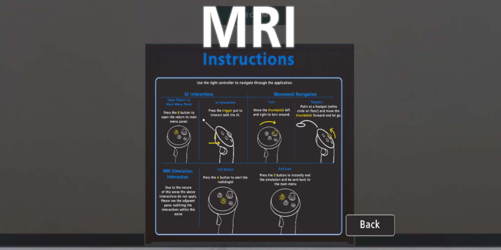
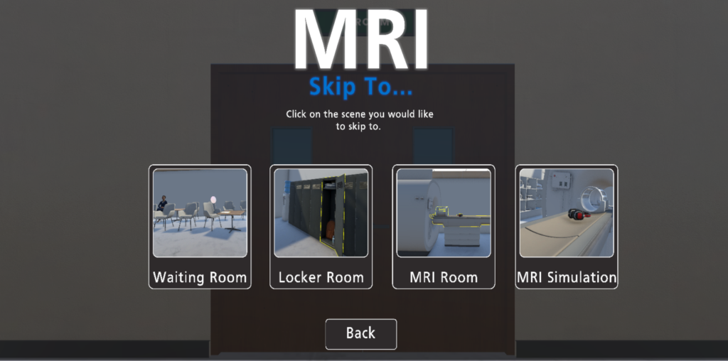
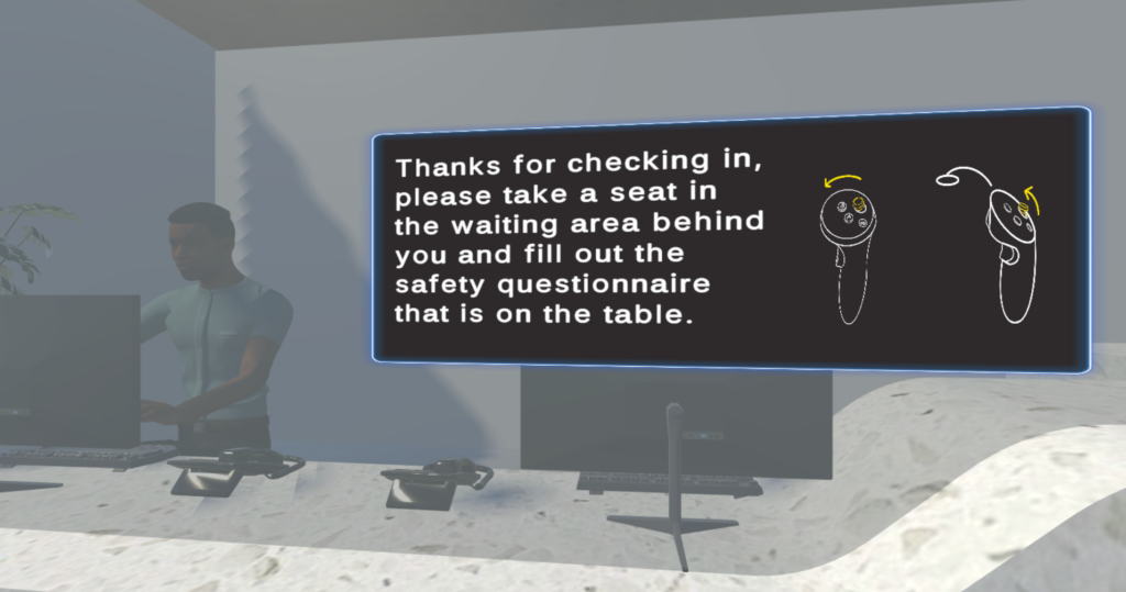
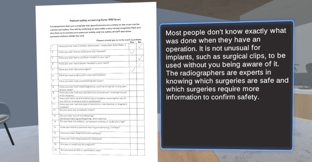
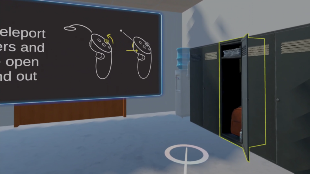
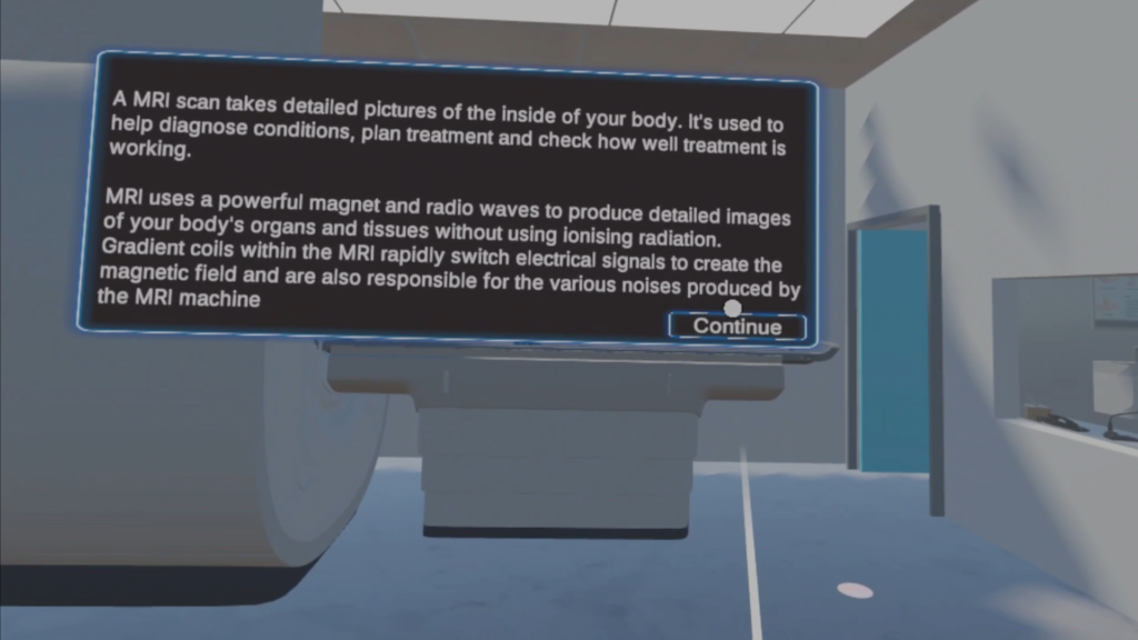
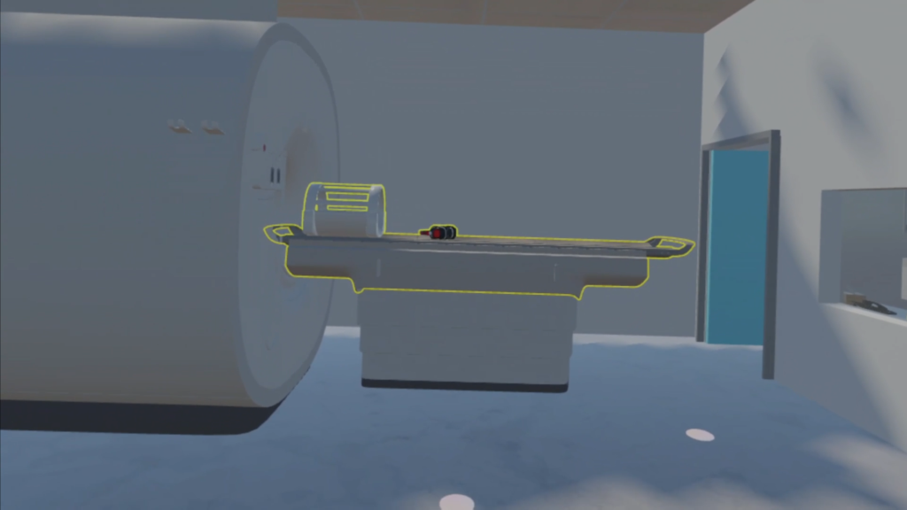
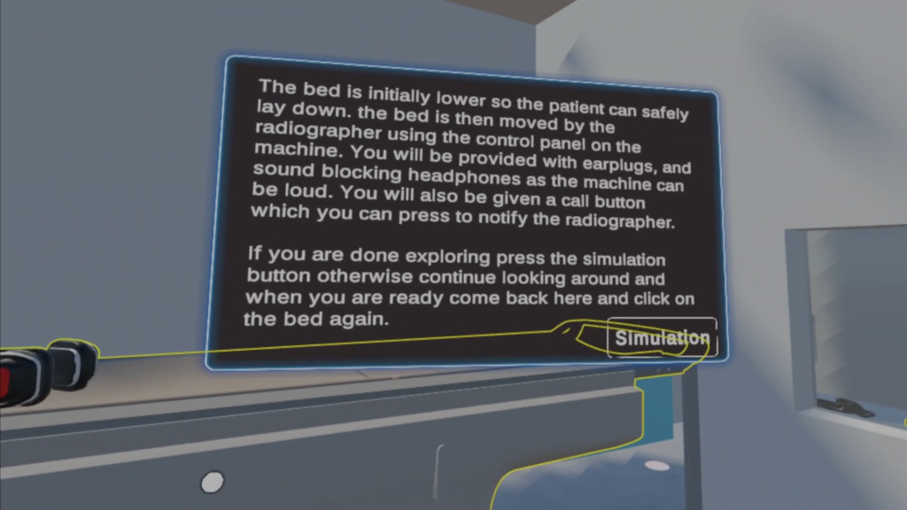
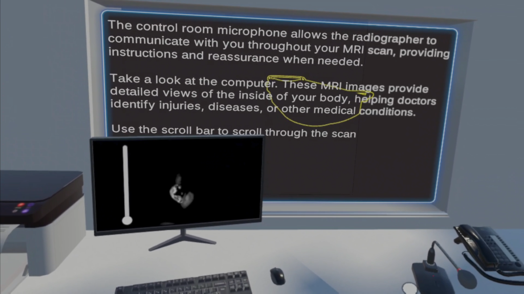
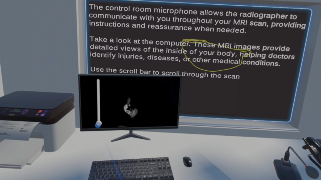
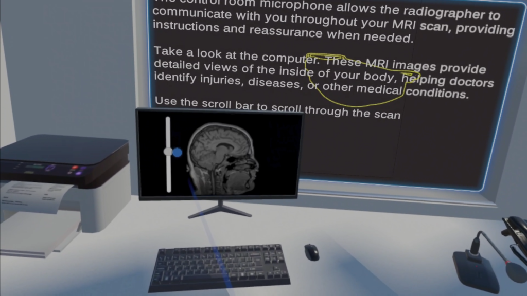
I designed, developed and evaluated an immersive virtual reality (VR) application intended to familiarise adults with the magnetic resonance imaging (MRI) scanning experience in collaboration with the Lancashire Teaching Hospitals NHS Foundation Trust (LTHTR), with the aim of helping users better understand what happens during an MRI examination and what the scanning environment may feel like.
Patients may feel anxious about magnetic resonance imaging (MRI) for various reasons, including the enclosed bore, loud noise, and the need to remain still during the procedure, which can affect the patient experience and may lead to premature scan termination, reduced image quality and, on occasion, cancellations. Existing interventions such as general anaesthesia or sedation introduce additional risks and higher costs due to equipment and increased staffing requirements. Traditional preparation methods, such as informational leaflets, cannot convey sensory and spatial information alone. VR offers an alternative approach to patient preparation by allowing users to experience a simulated MRI environment and procedure before undergoing a real examination. Rather than just providing information VR can introduce users to aspects like the scanner environment and sounds, experience within the bore, patient positioning and the requirement to remain still.
This study followed a structured design and development process, including observational visits and professional input, existing research, and NHS guidance, to identify key requirements of the MRI patient journey. These requirements were then translated into design specifications and developed into an interactive VR application for the Meta Quest 3 using Unity. The application was then evaluated with participants from GSA and the public. The experiment assessed measures such as MRI-related anxiety, preparedness and knowledge, presence, and usability using a mix of validated and bespoke questionnaires, and the results showed evidence that the VR experience improved patient understanding, preparedness, and confidence while reducing anxiety. The application also demonstrated a high sense of presence and usability.
Overall, the project explored how immersive VR could provide both educational and experiential familiarisation for MRI, allowing users to experience some of the spatial and sensory aspects of the examination. This prototype provides a foundation for further development and evaluation within the clinical setting.
3D Modelling & Animation
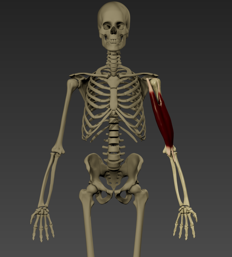
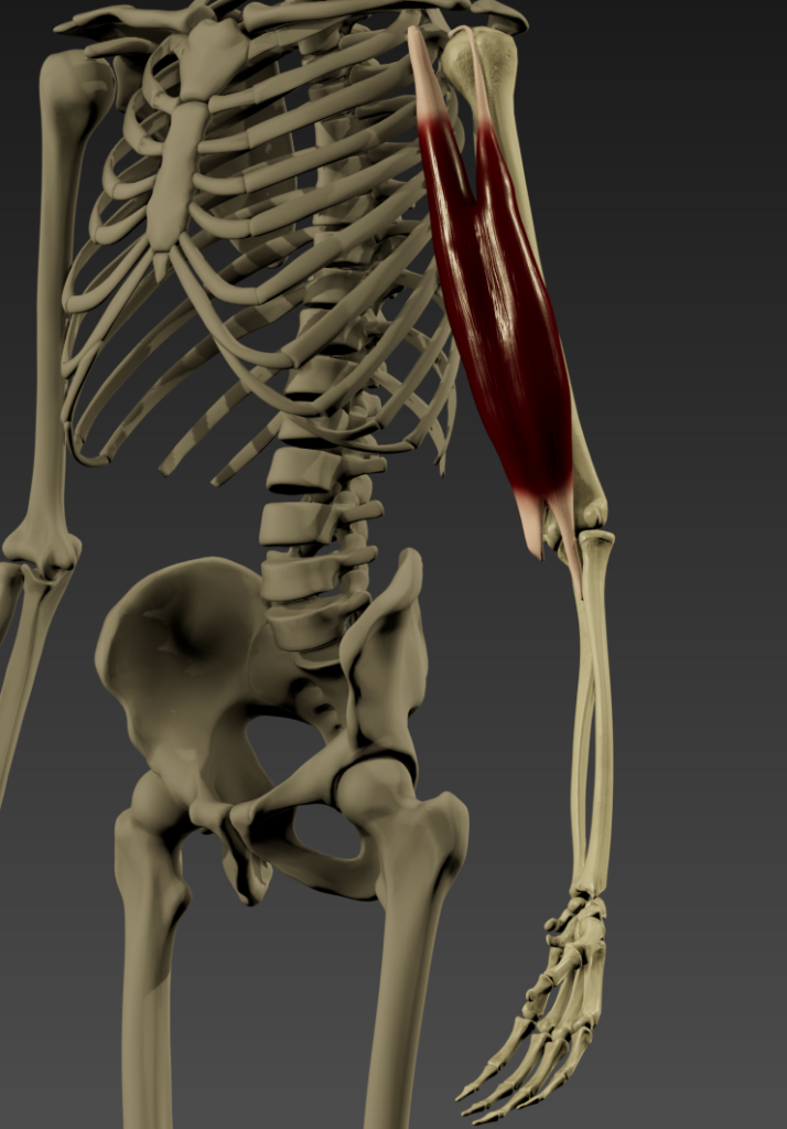
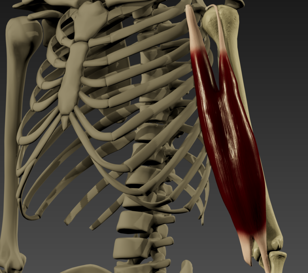
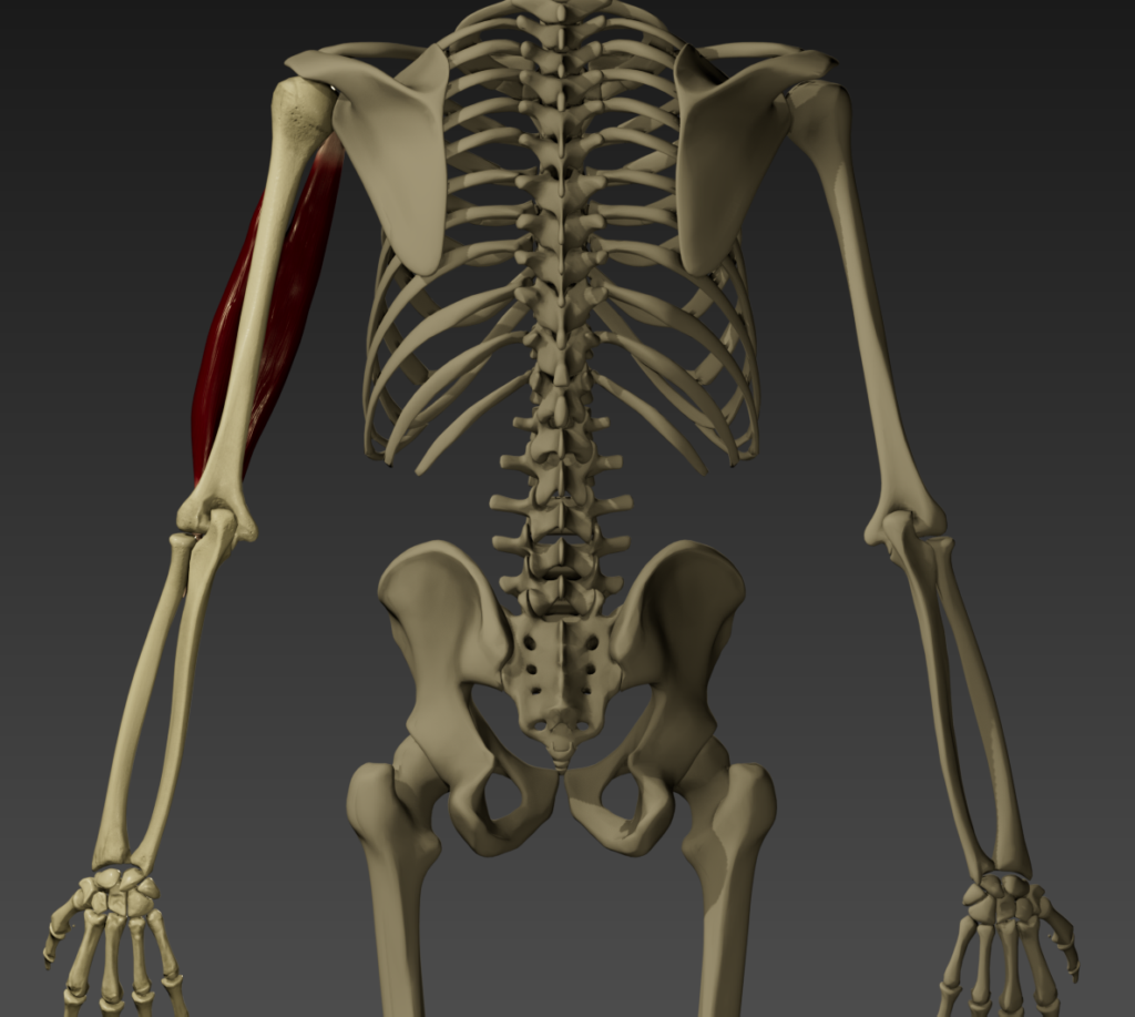
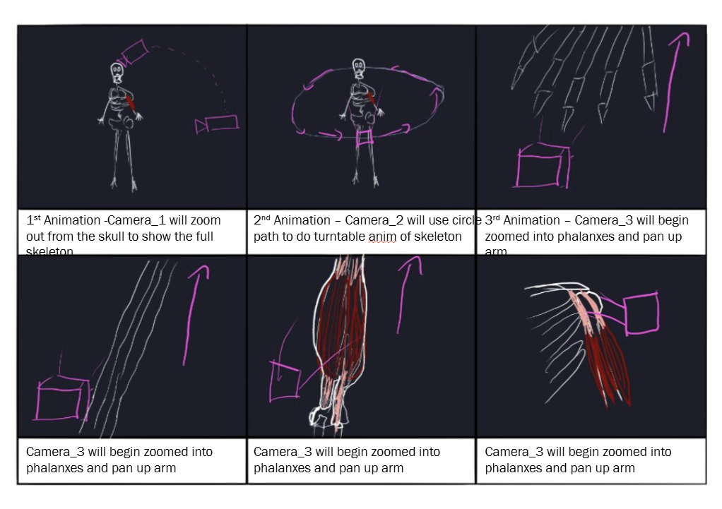
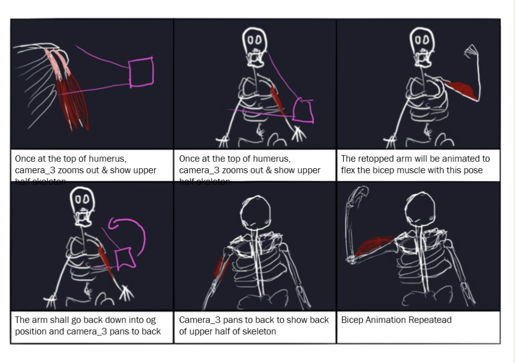
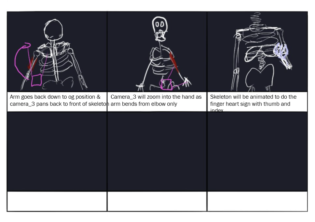
This project involved using software such as 3ds Max and ZBrush to retopologise the arm bones, model a bicep from scratch, and animate it all using morphers for a final visual outcome.
Visualising Anatomy – Volumetric Visualisation
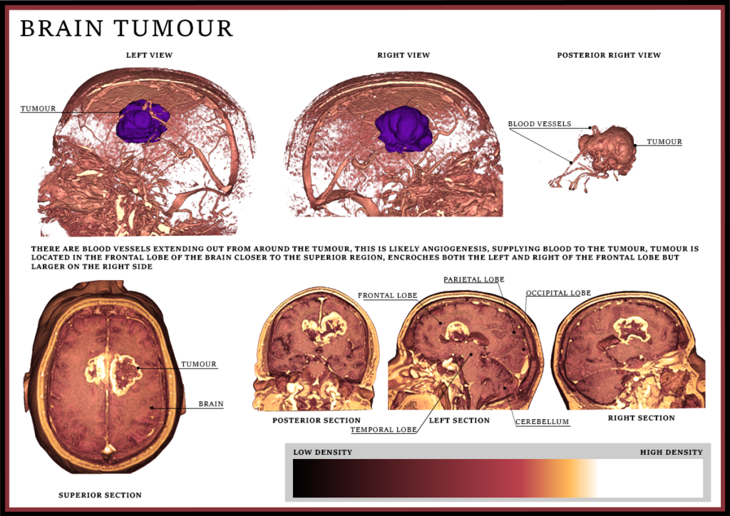
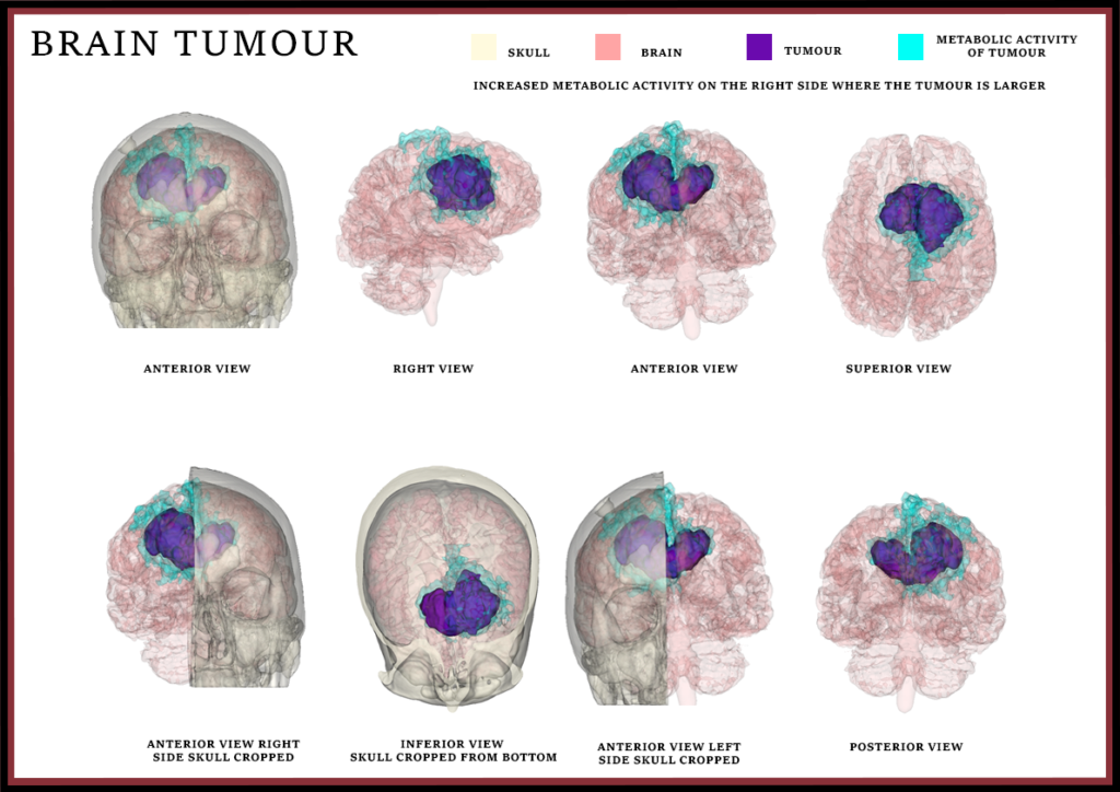
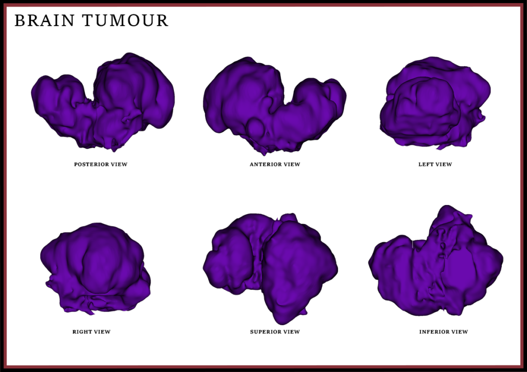
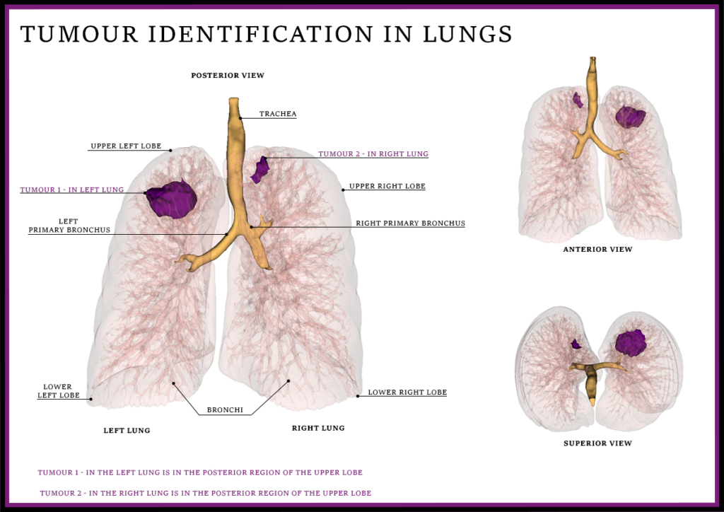
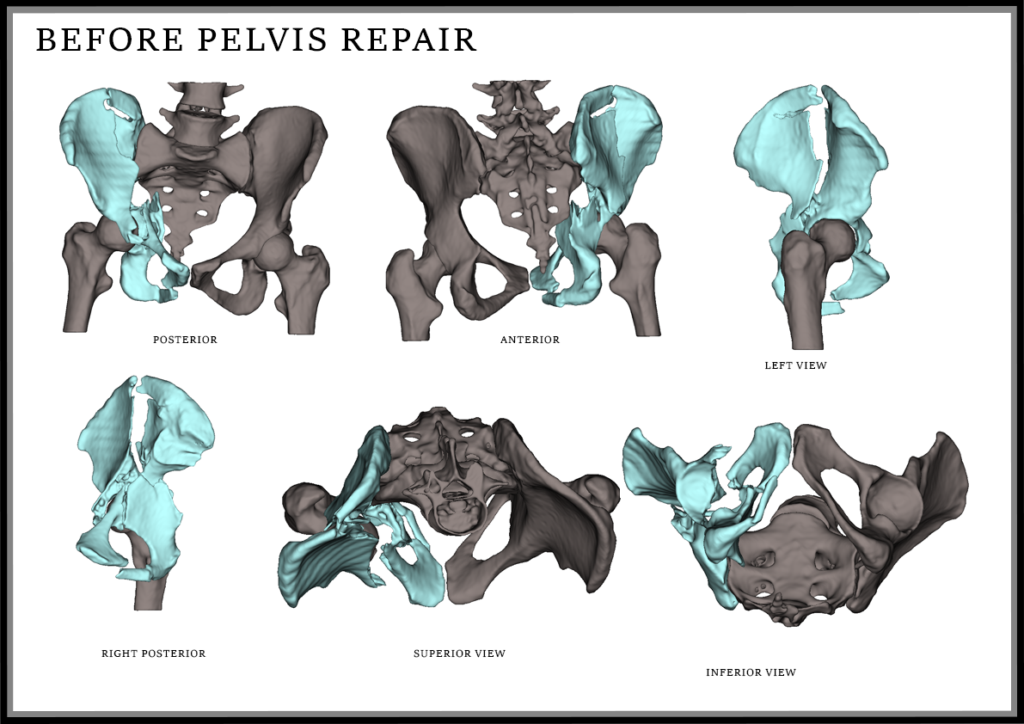
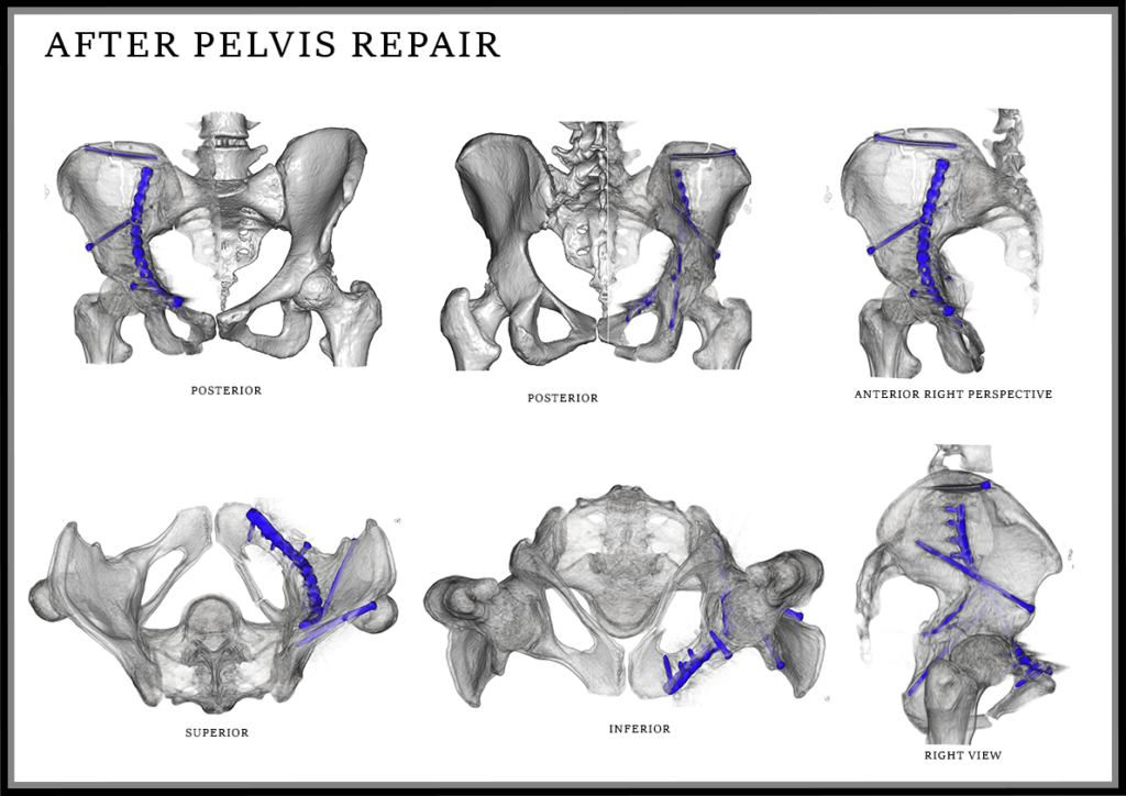
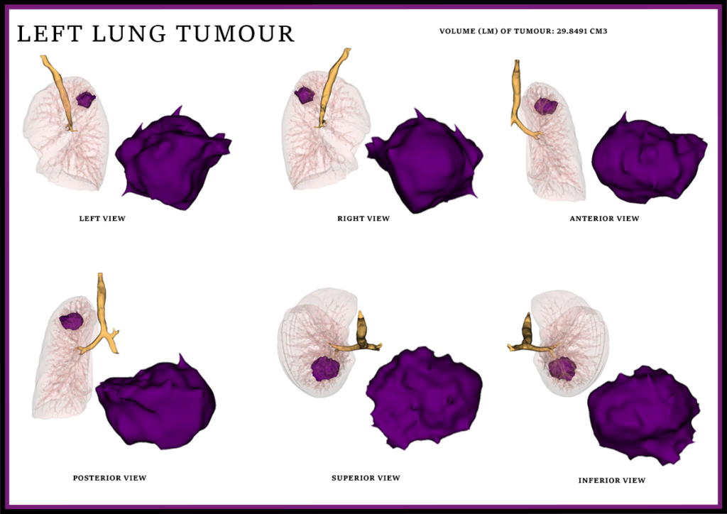
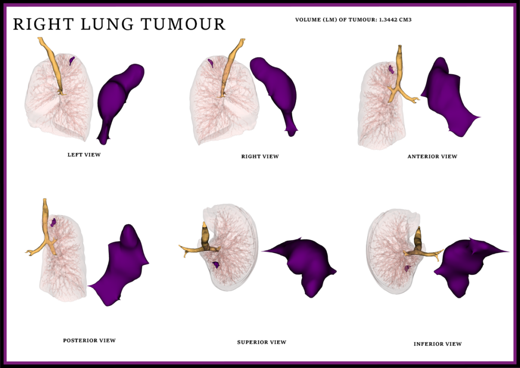
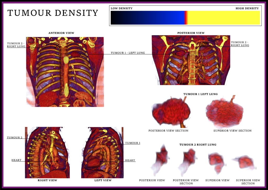
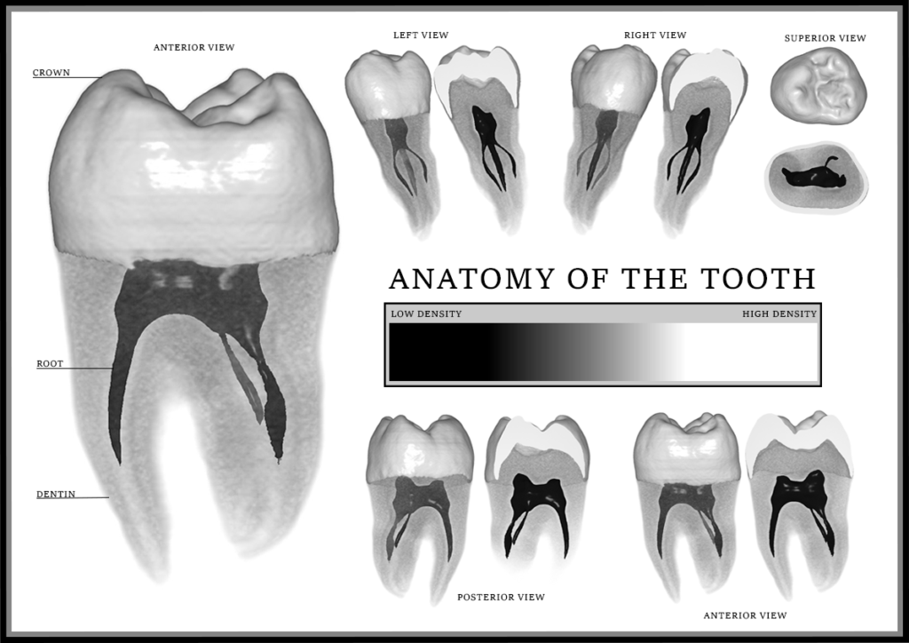
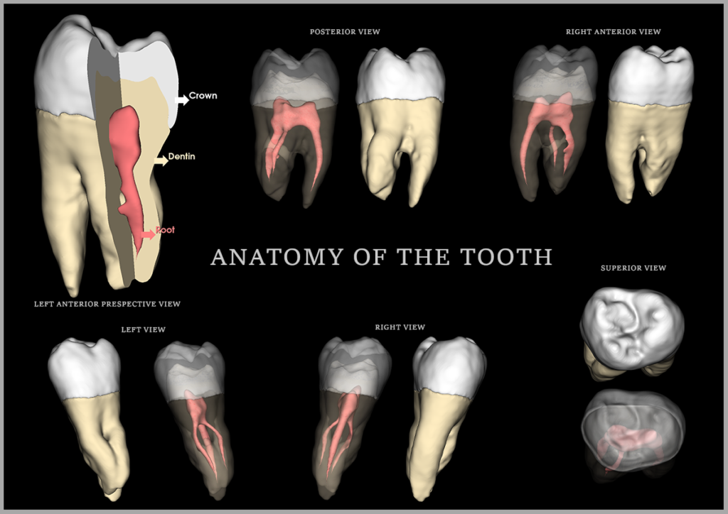
Datasets from MRI, PET, and CT scans were imported into 3D Slicer and MITK, then developed into 3D visuals using direct and indirect rendering techniques.