Wei Lin
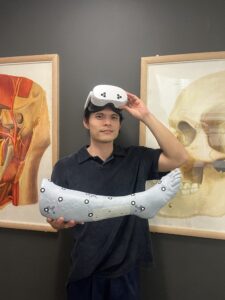
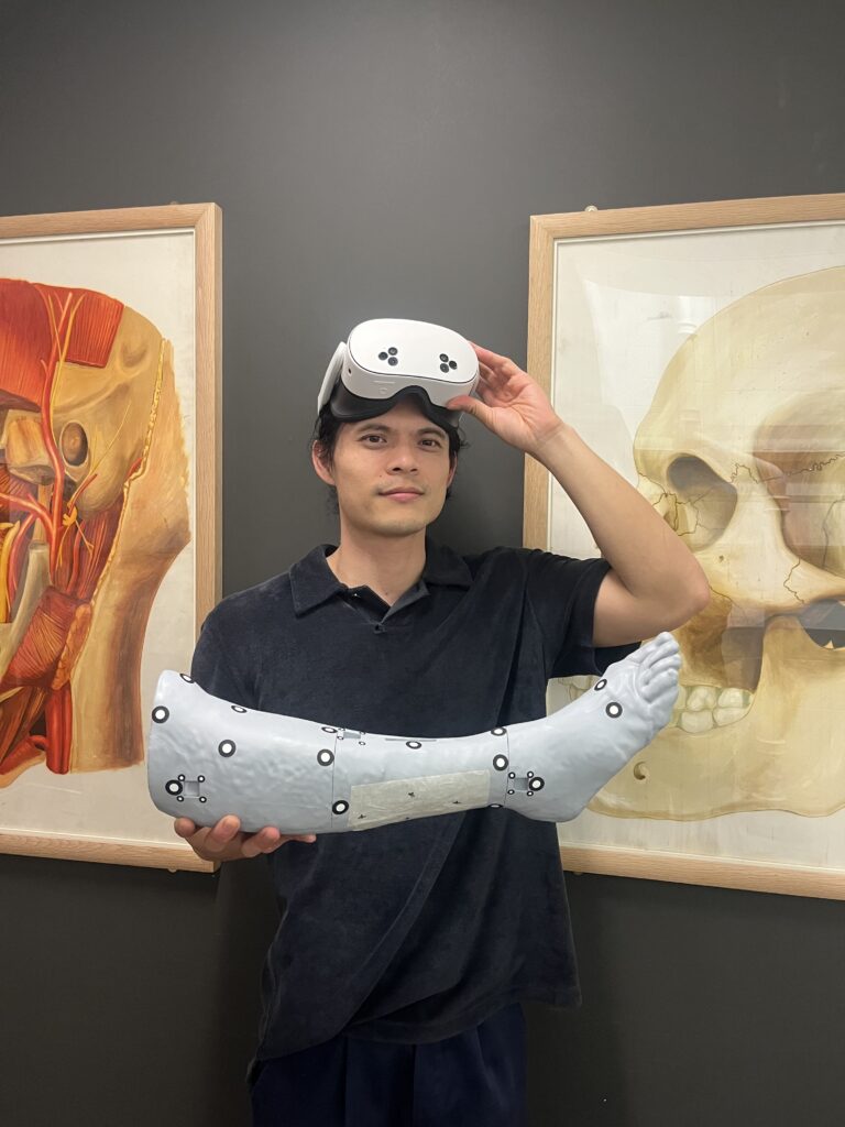
Wei Lin is a multidisciplinary medical visualisation practitioner working across human anatomy, digital modelling, physical fabrication and extended reality. During his MSc in Medical Visualisation and Human Anatomy, he developed experience in medical image processing, anatomical 3D modelling, 3D printing, 3D scanning and XR-based surgical navigation research. His final research project investigates how accurately the Meta Quest 3S can overlay planned free fibula flap margins onto a 3D-printed leg model, combining 3D Slicer, 3ds Max, Unity, QR-code-based spatial registration and Artec scanning.
Before specialising in medical visualisation, Wei worked in architectural design, where he developed a strong foundation in CAD, spatial problem-solving, multidisciplinary collaboration and technical communication. His wider creative practice also includes architectural and ceramic design work presented in exhibitions. These experiences have shaped his interest in bringing together anatomy, technology, material making and spatial design to communicate complex medical ideas and explore their potential applications in healthcare.
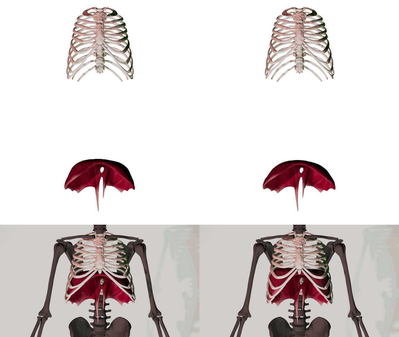
Modelling and Animation of the Rib Cage and Diaphragm
Modelling and Animation of the Rib Cage and Diaphragm
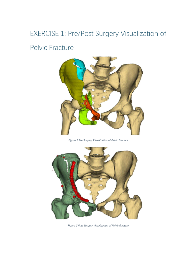
Volumetric Visualisation

Evaluating the Registration Accuracy of Meta Quest 3s Mixed Reality for Free-Flap Surgery Overlay on a 3D-Printed Leg Model
This project was conducted in collaboration with the University of Aberdeen and the MSB Medical School Berlin, and I would like to thank everyone who contributed to or took part in this study. I would also like to express my gratitude to my supervisors, Dr Matthieu Poyade, Prof. Flora Groening, Dr Jenny Clancy, and Dr Jenny Gregory and our collaborator Mr Terry Lowe, Consultant Maxillofacial Head and Neck Surgeon at the Aberdeen Royal Infirmary, for their continuous support and guidance throughout the project.
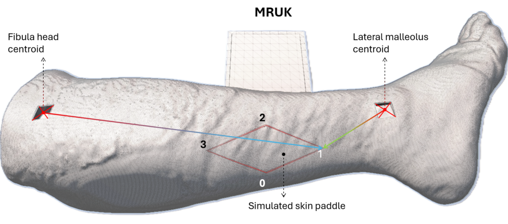
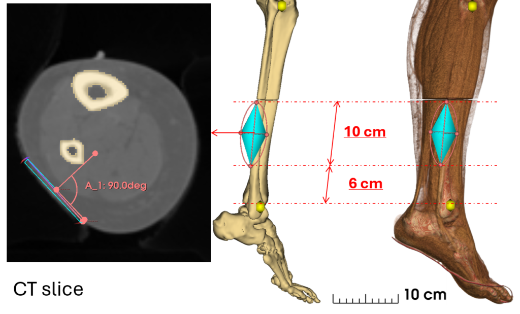
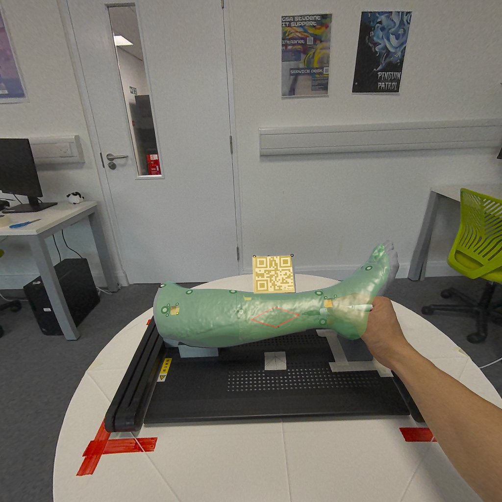
This project investigated whether a Meta Quest 3S mixed reality headset could accurately position a virtual surgical plan on a physical 3D-printed leg model. The study was based on free fibula flap surgery, with the planned margins of a skin paddle displayed as a virtual overlay on the leg surface.
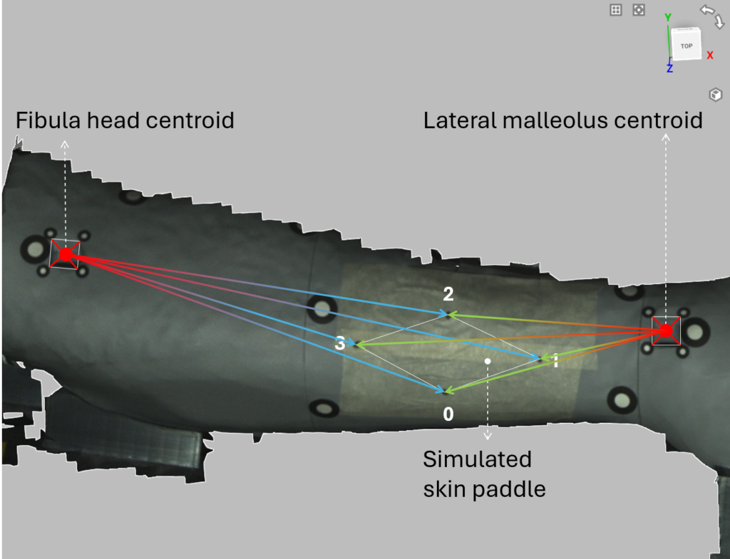
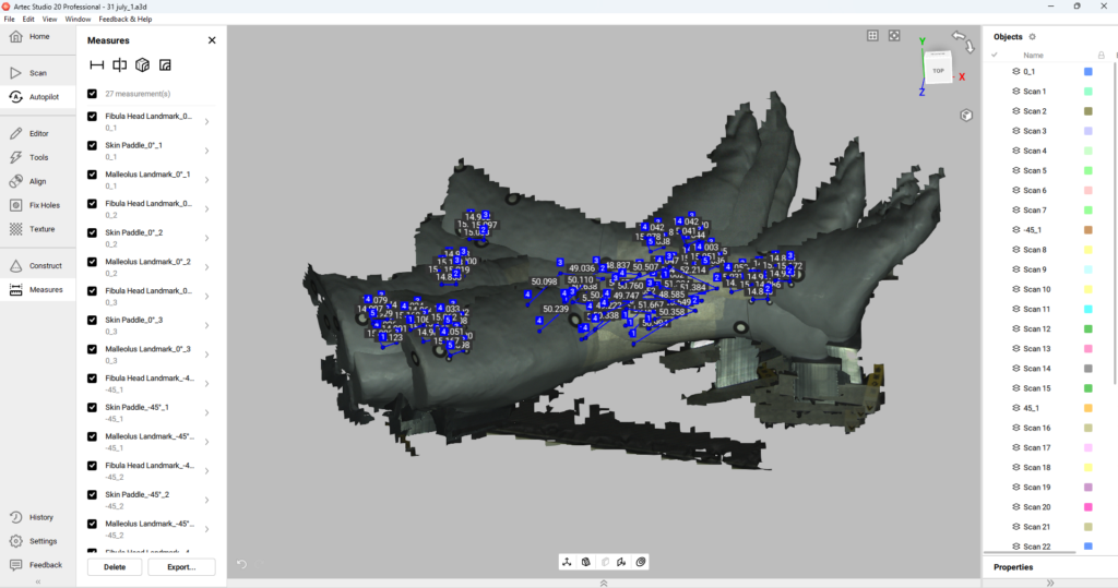
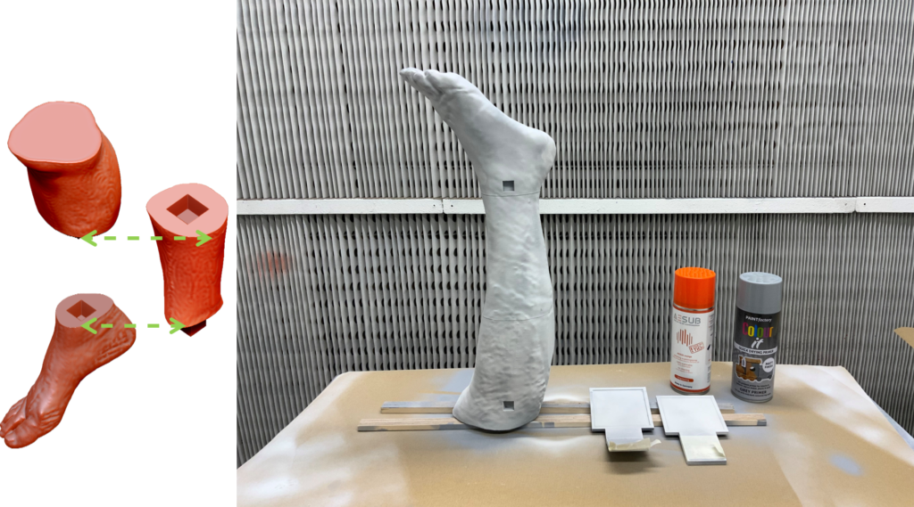
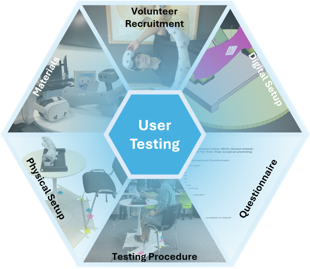
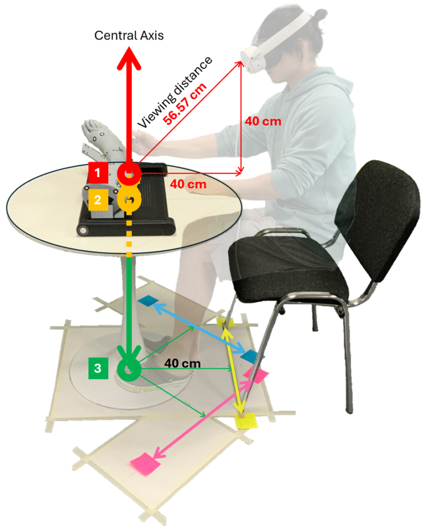
The overlay was developed in Unity using two QR-code-based approaches: the Mixed Reality Utility Kit (MRUK) and AR Foundation with OpenXR Spatial Entities. Participants viewed the overlay from three different angles and marked its four endpoints on the model. These markings were captured using an Artec 3D scanner and compared with the original digital plan using anatomical reference landmarks and a distance-based evaluation method.
By examining the effects of development pathway, viewing angle, anatomical reference location and flap endpoint, the study assessed the feasibility and limitations of consumer mixed reality for transferring surgical plans onto physical anatomy. The project brought together medical image processing, 3D modelling, additive manufacturing, XR development, 3D scanning and quantitative evaluation.
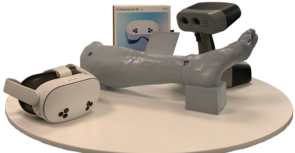
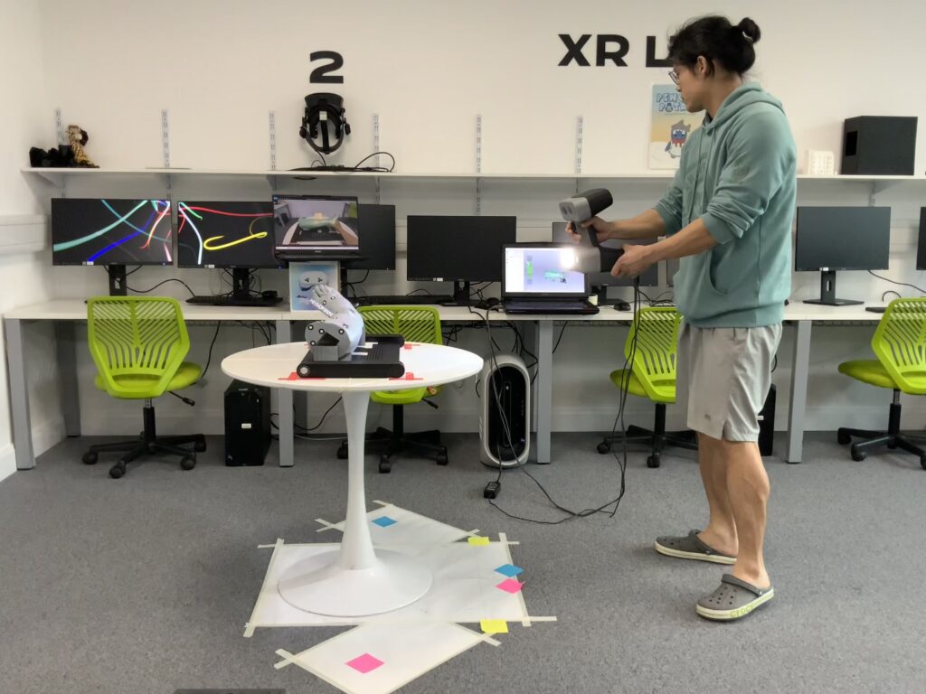
Scanning process