
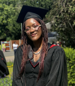
Precious Karley
(she/her)
My name is precious Karley, and I’m an illustrator at heart, with a background in law. During my studies I was fascinated by the institutional structures in place that maintain our relationship as individuals with the healthcare system. Medical negligence and criminal cases gained my interest as I enjoyed hearing expert witness testimony from clinicians. Visuals are often used to communicate complex pathology and injuries to the jury. Representing these diagrammatically through illustration is an excellent way of decreasing ambiguity and facilitating top down communication. Information is always received depending on the perceptions of the audience, and the application of art to the field of medicine provides opportunities for opaque concepts to be communicated effectively.
My interest in anatomy began as I was a painter in high school, trying to perfect basic consistent forms of the human body. I studied the works of Renaissance painters, fascinated by their cadaveric dissection for the purposes of visual understanding. Since then I always drew with the underlying structures in mind, recreating the musculature beneath the skin to approach anatomical accuracy.
I have always found medical illustrations insightful and beautiful. For me, they are a reminder of the evidence of design in the human body. Specific ranges of motion to adhere to, with organic shapes in the form of repeating patterns. As someone without a formal scientific background, I’ve always had the desire to receive clinical education to deepen my understanding. With the aims of gaining functional knowledge of the human body to aid the accuracy of my illustrations, as well as the opportunity to contribute to the field of scientific communication. Here at Glasgow School of art, I’ve had my creative potential nurtured. This year, it felt like my dreams came true.
I have had the opportunity to dissect cadavers and gain functional understanding of the human body. I have developed 3D modelling skills, C# programming, game development and volumetric visualisation.
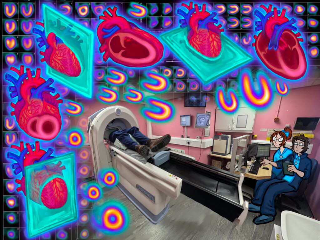
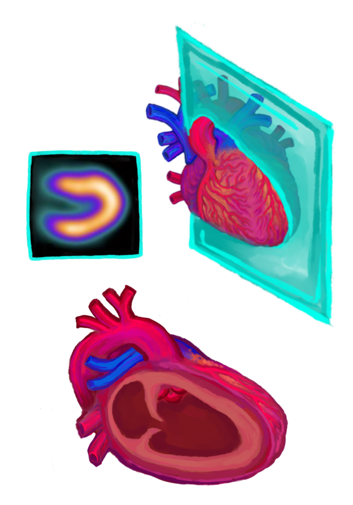
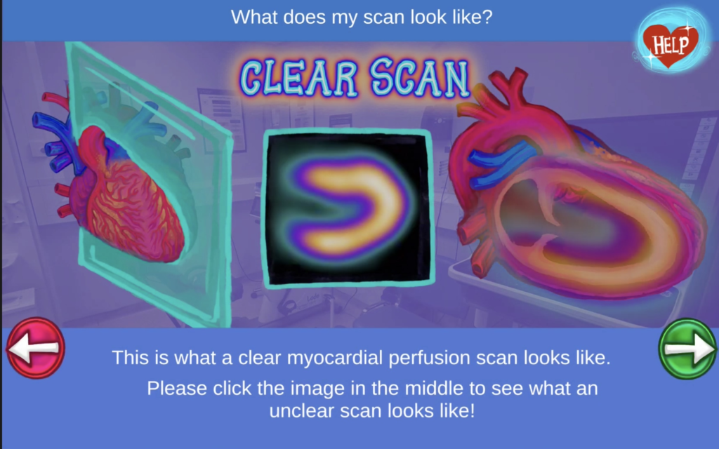
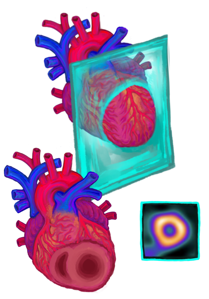
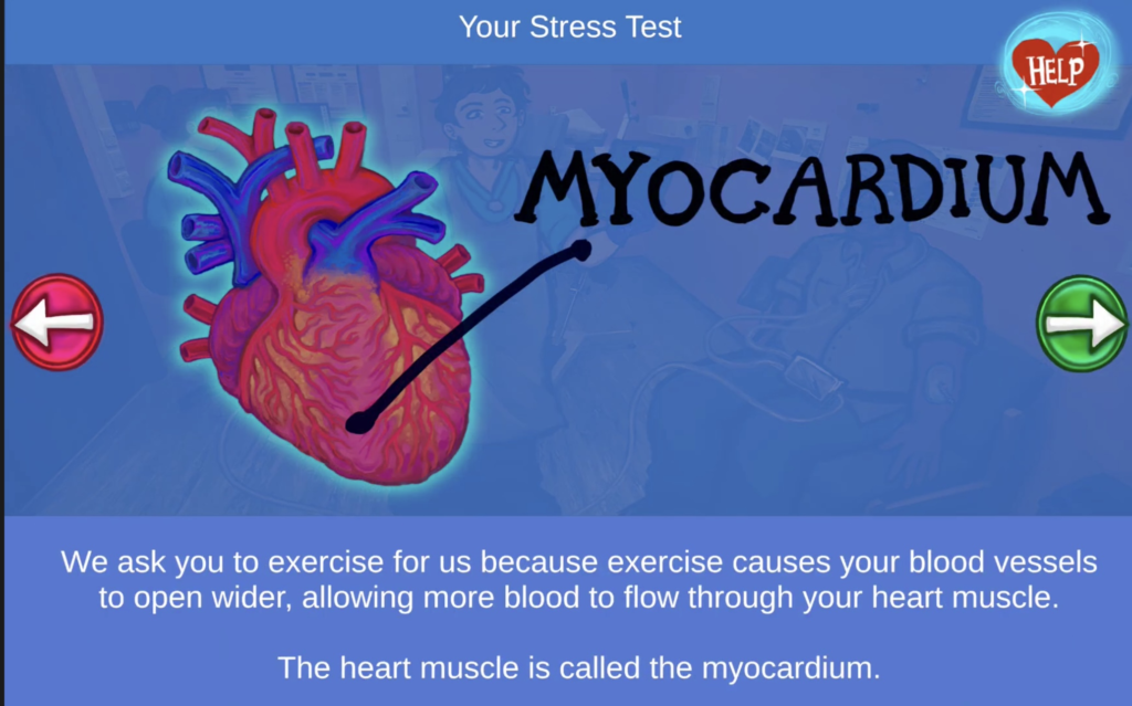
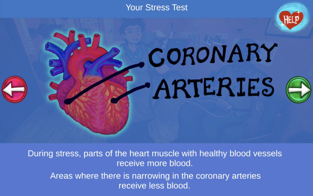
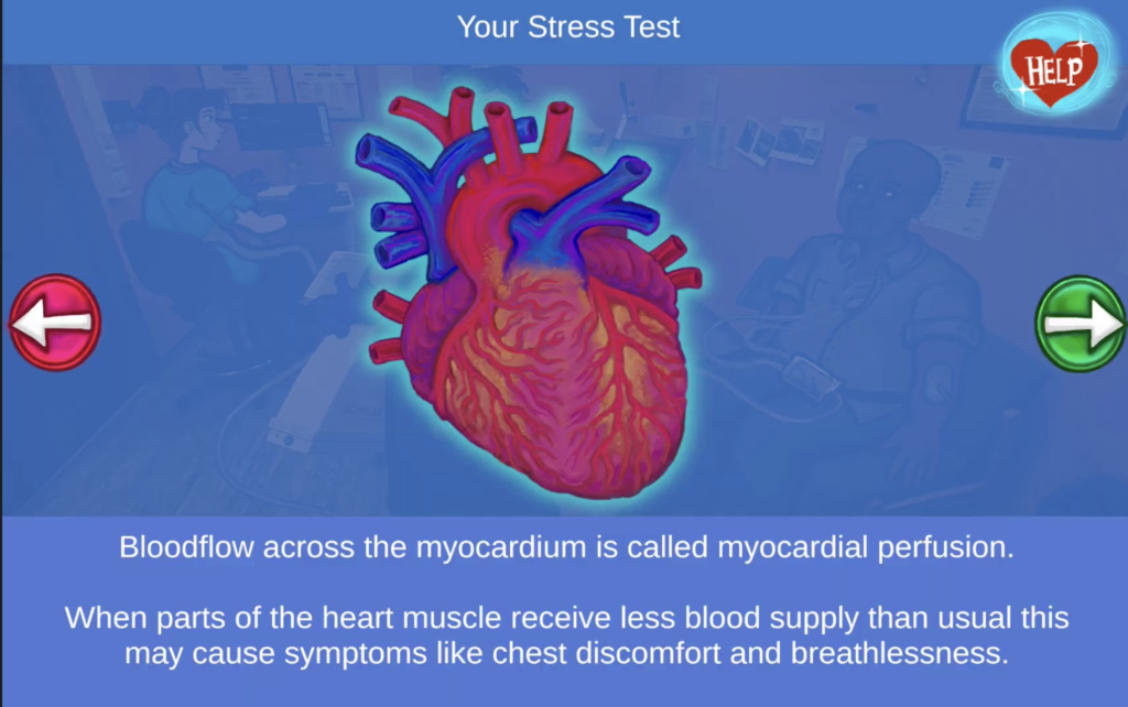
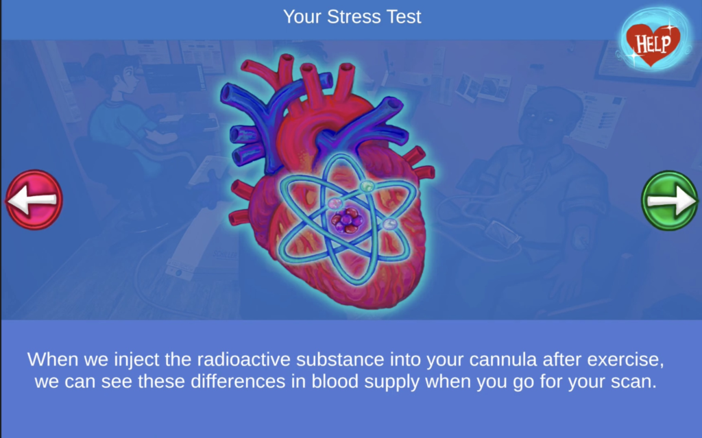
THE SCAN: Evaluation of a Patient-Centred Multimedia Application
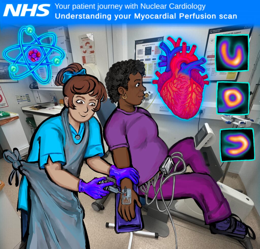
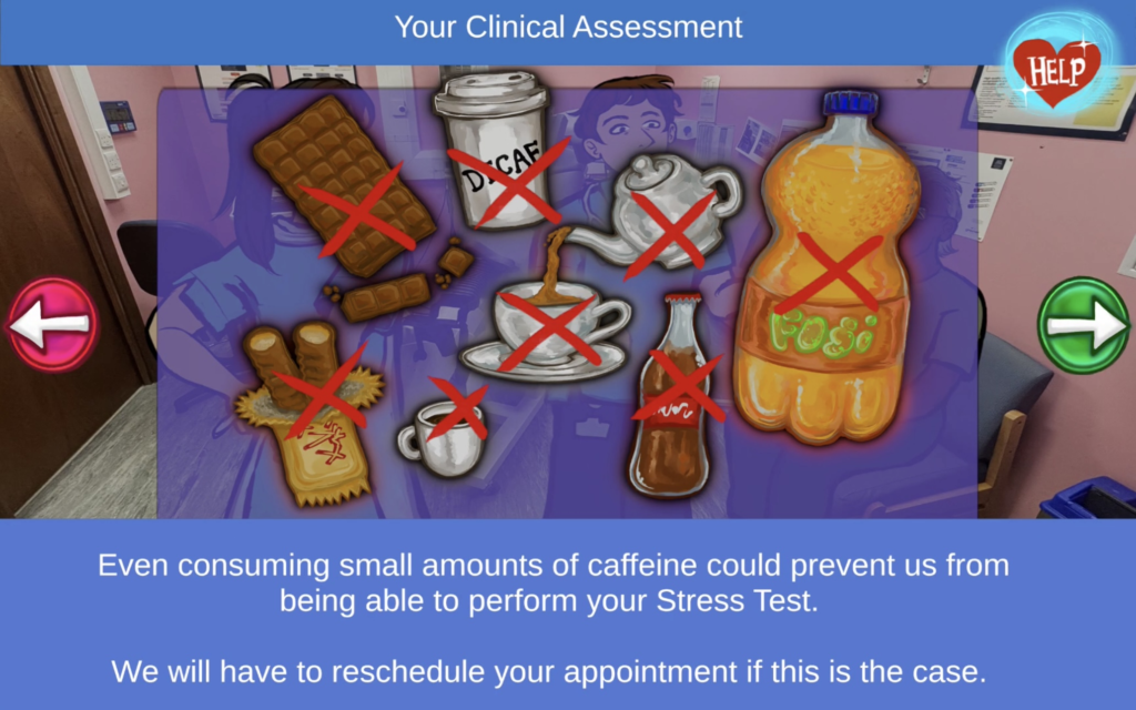
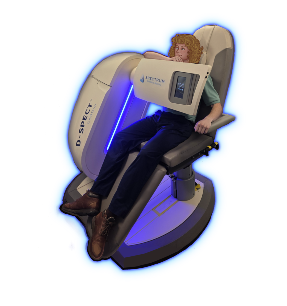
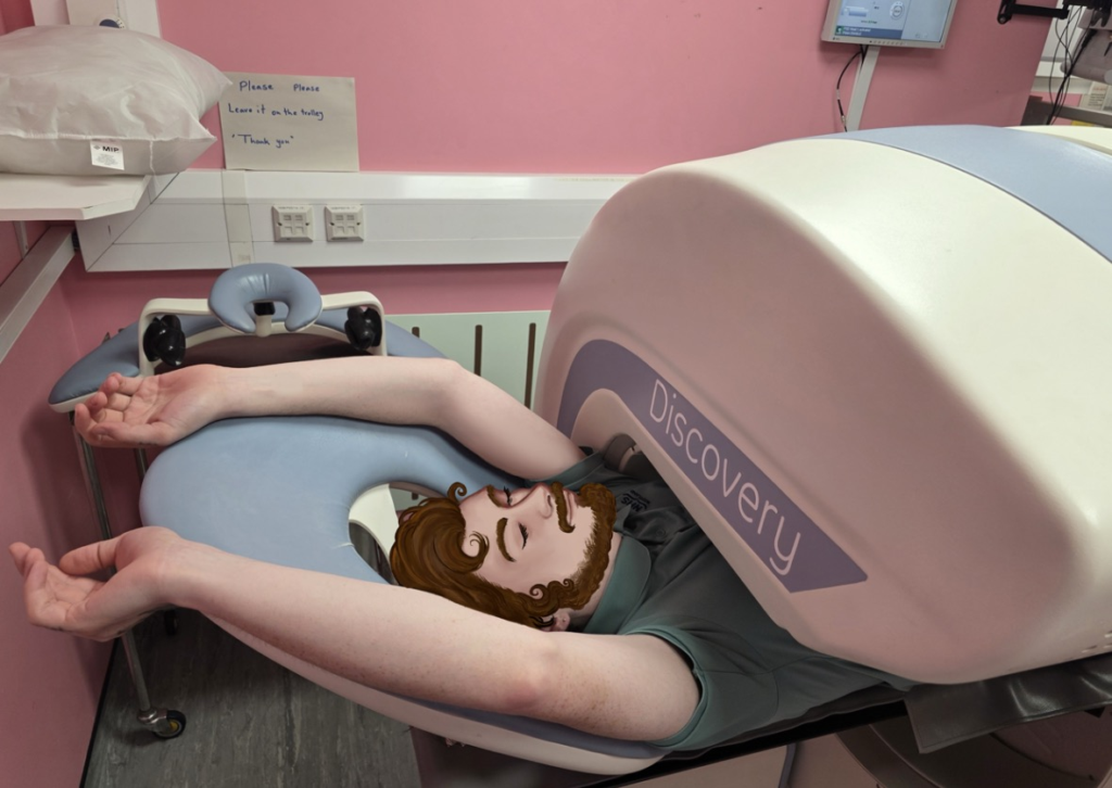
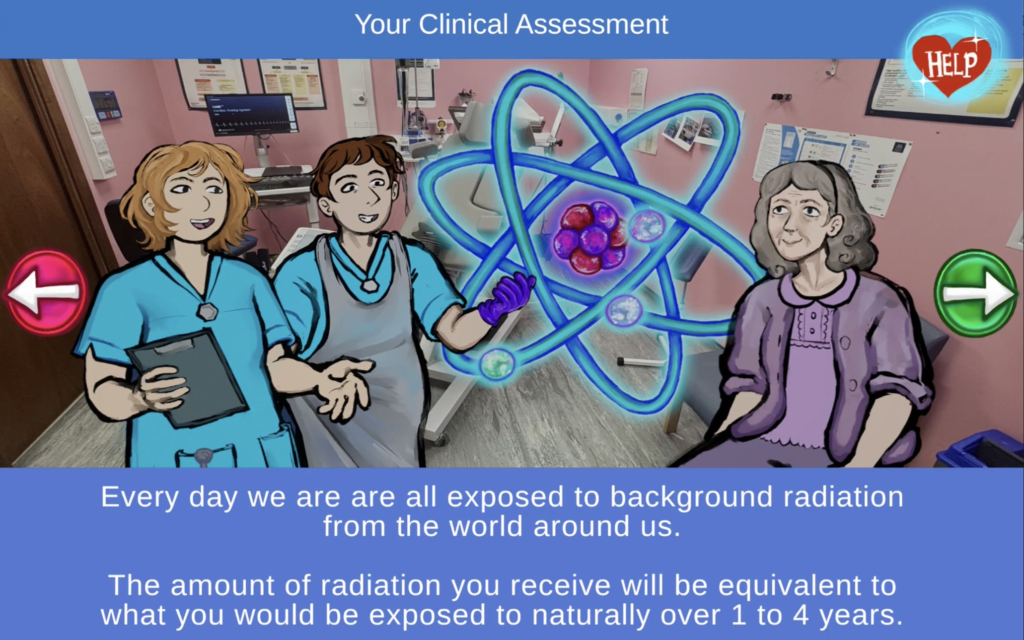
Patients attending Nuclear Cardiology departments for myocardial perfusion imaging often arrive with negative expectations and misconceptions about their appointment experience. Patients undergoing cardiac imaging are often struggling with chronic illness and existential uncertainty. Although leaflets deliver the appropriate clinical information about their appointments, they don’t serve the prescient need for reassurance.
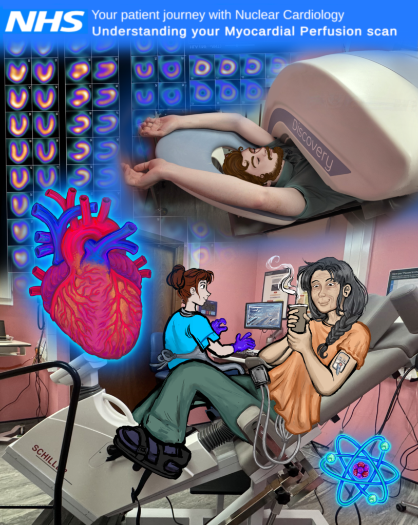
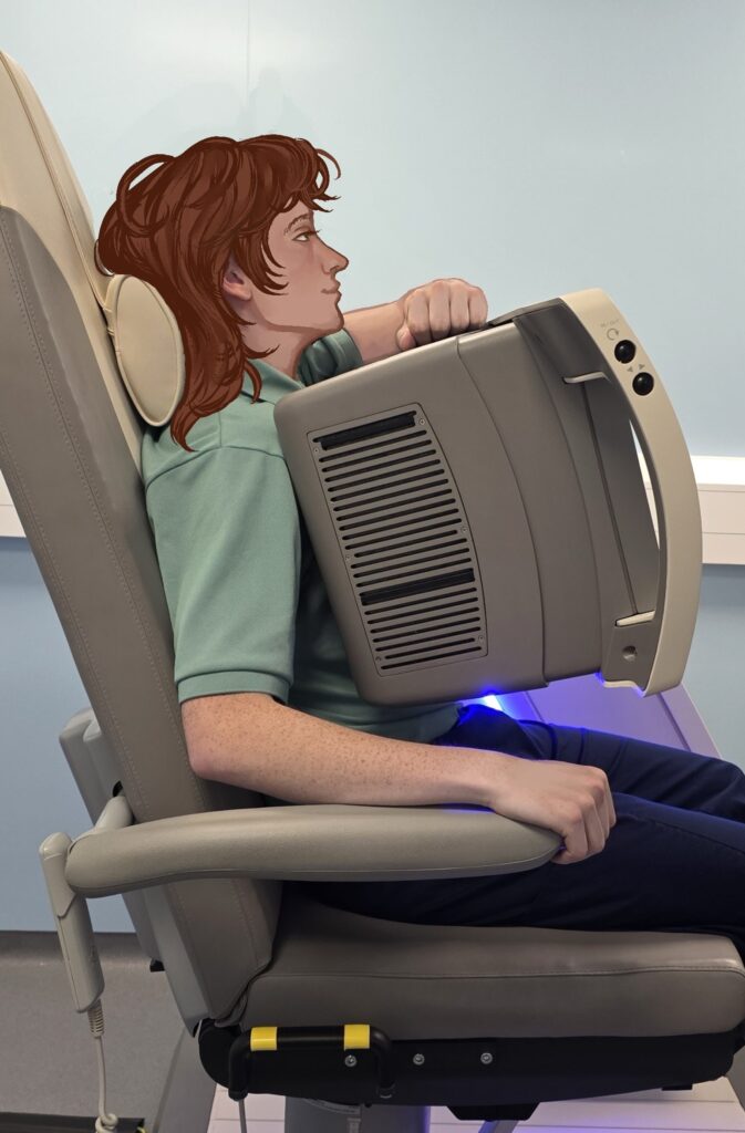
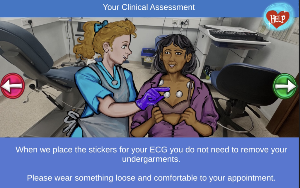
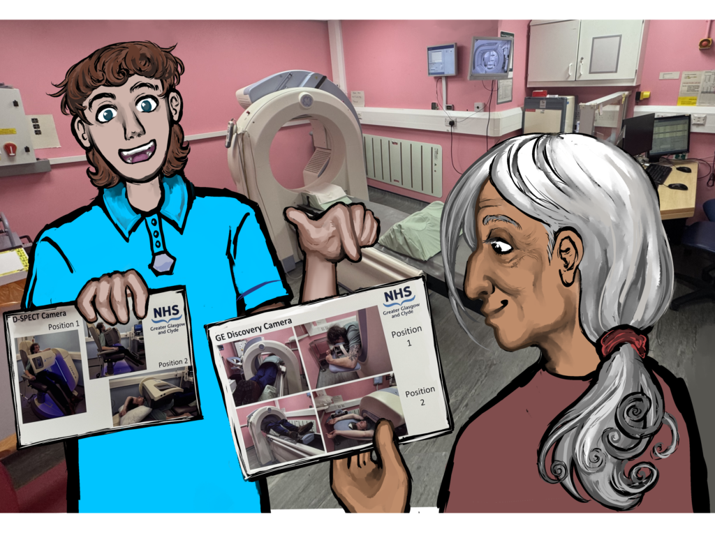
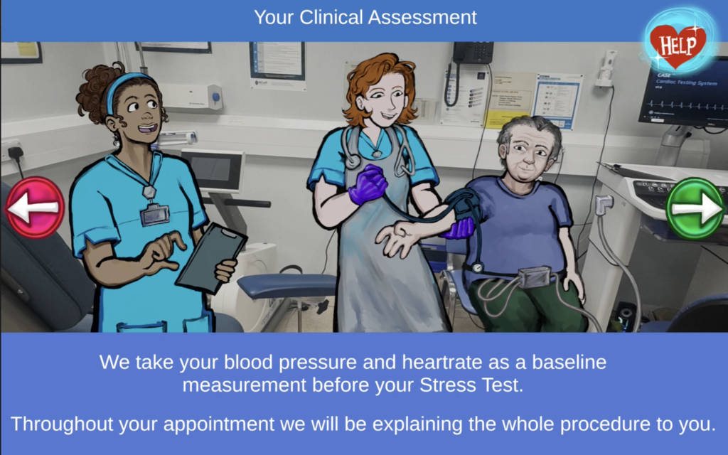
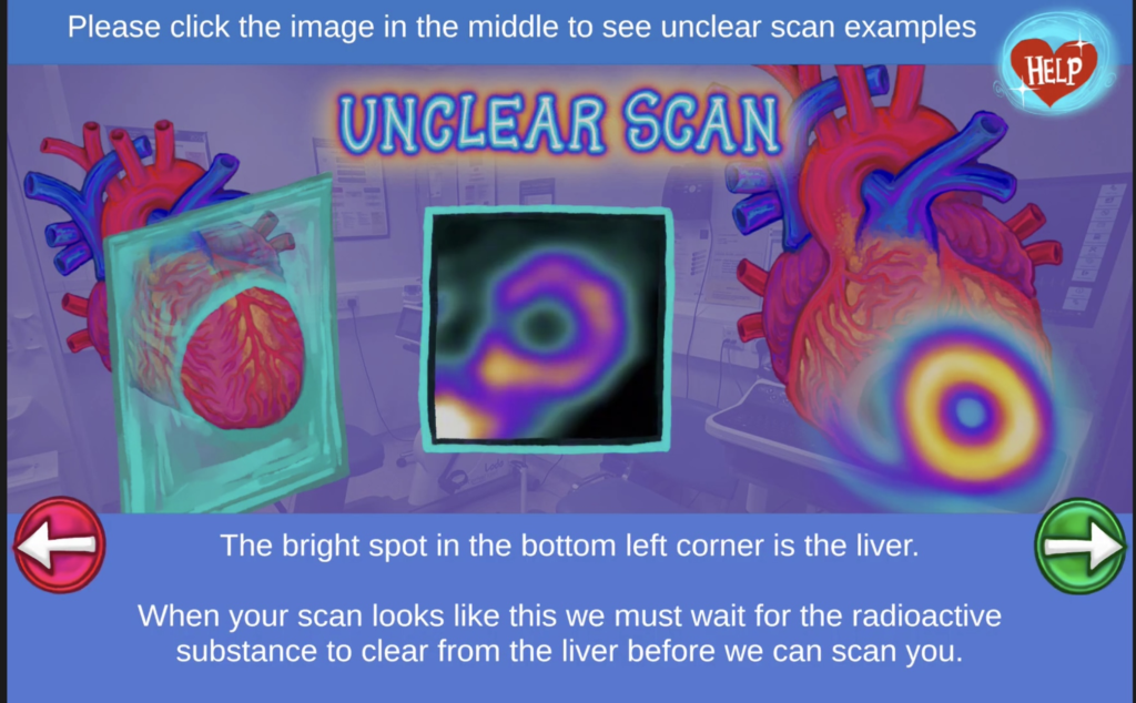
This mobile Application was developed in collaboration with NHS Greater Glasgow & Clyde. With this development, I utilised the psychoeducational effect of narrative to deliver information to patients attending MPI appointments.
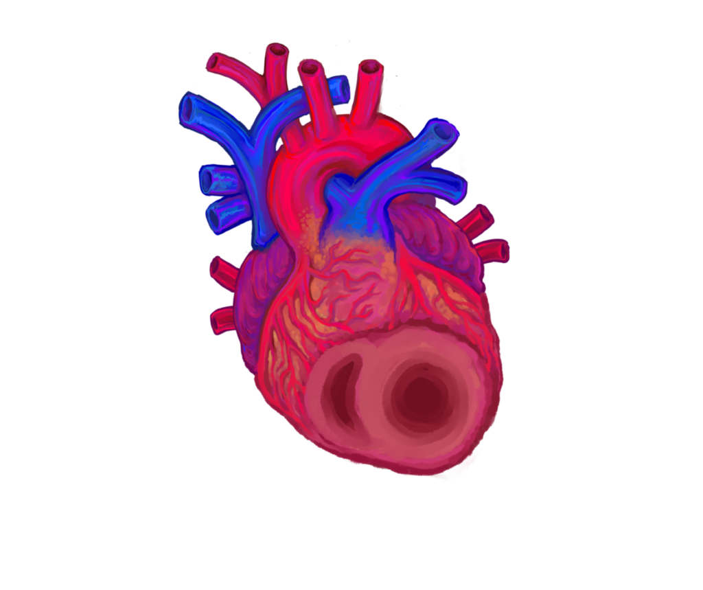
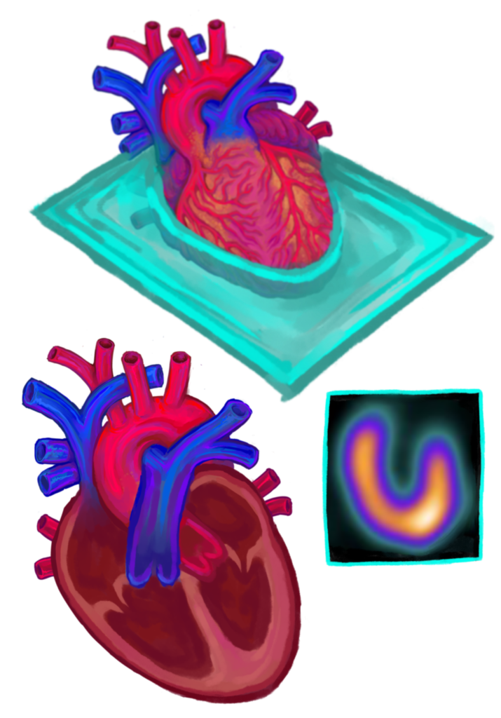
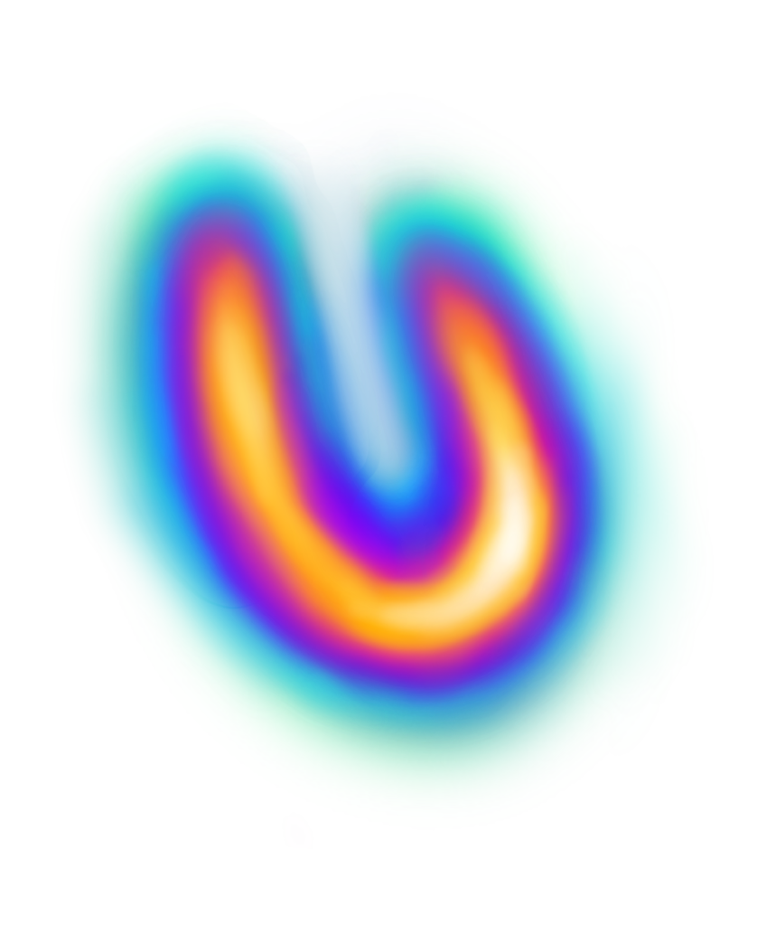
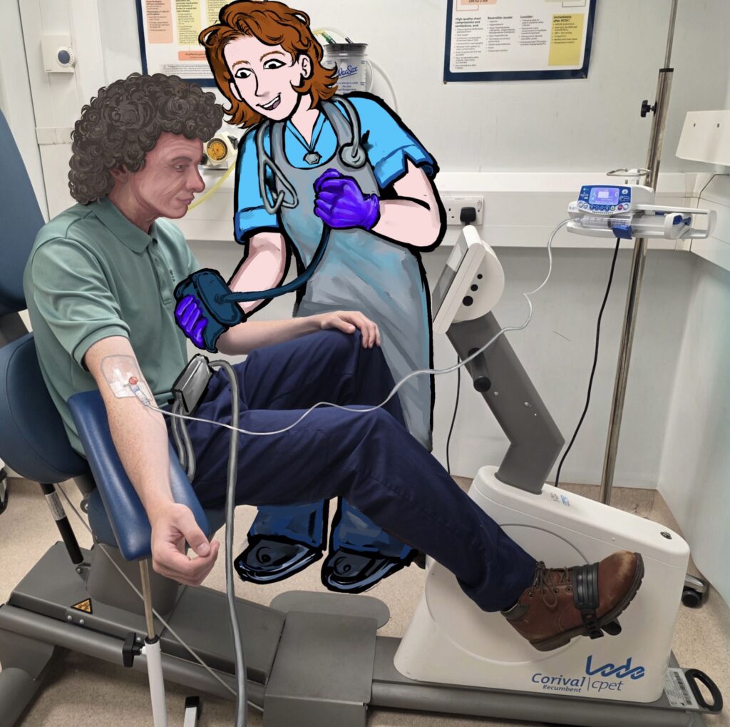
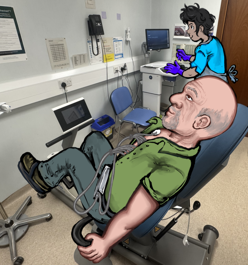
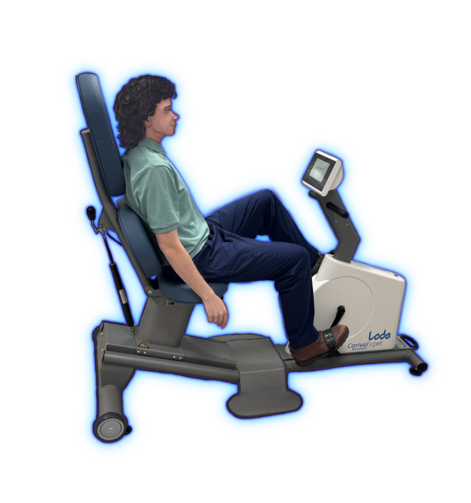
During 3 months of clinical observation I witnessed the compassionate, service of the Advanced Technologist team. Through life drawings and gesture based sketches, I captured realistic depictions of the patient experience for the purposes of patient centred education.
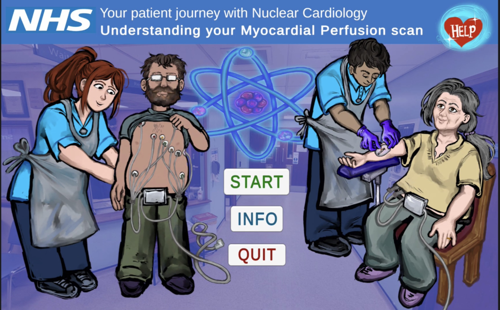
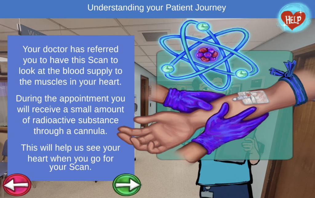
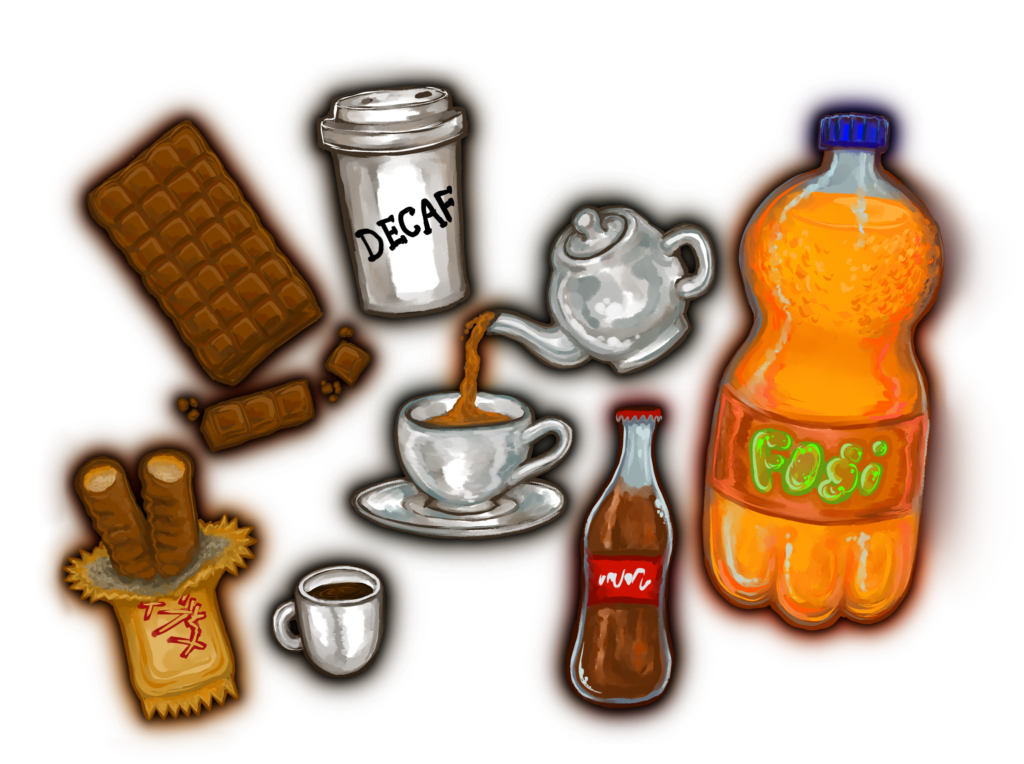
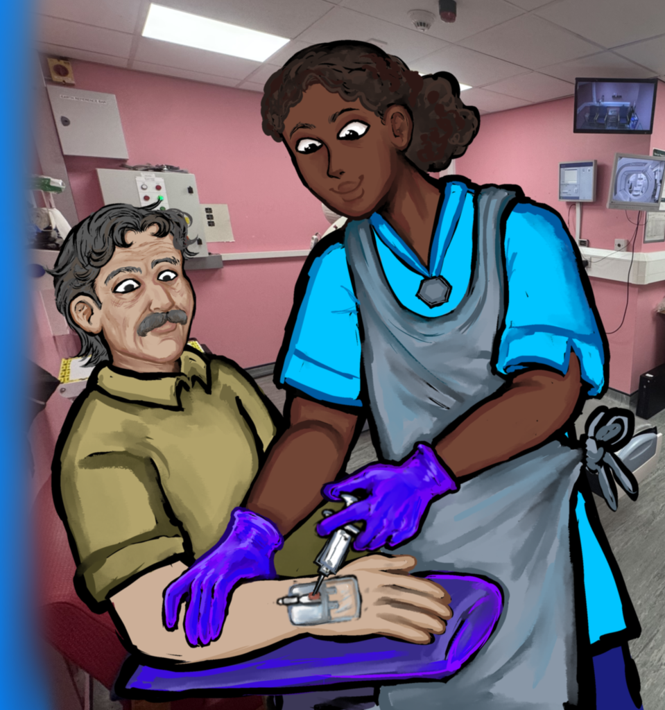
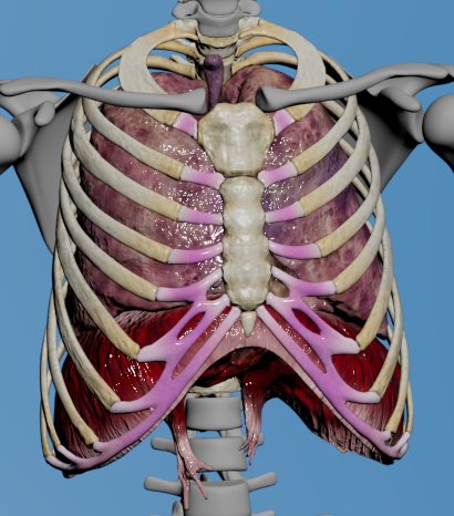
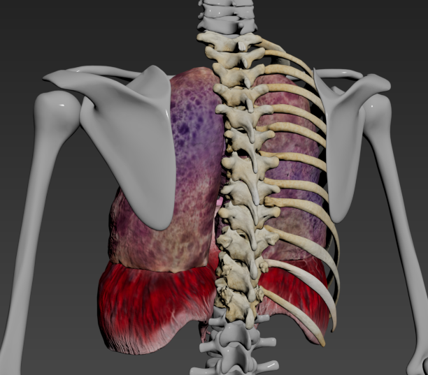
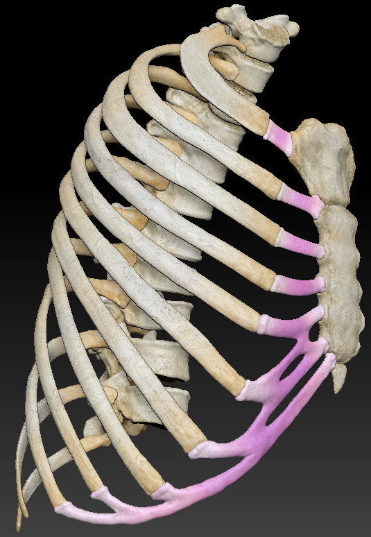
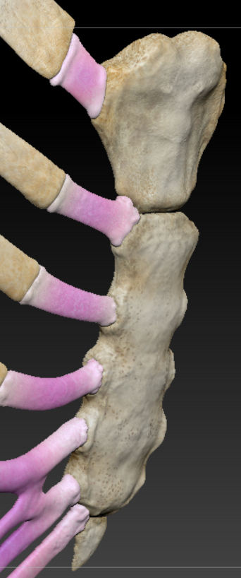
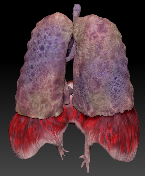
3D Modelling project: Diaphragm
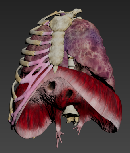
This assignment required us to retopologizse all 12 rib bones, The manubrium, Sternum, Xiphoid process and Vertebrae T1-T12. I additionally created new linkages in the coastal cartilage joining the ribs to the sternum to give the ribcage an organic look. Medical Visualisations usually give the cartilage a blue look to make it appear distinct from the surrounding bones, I personally chose to have my cartilage be pink because I thought it was more visually appealing than blue.
I modelled the Diaphragm from scratch. For the lungs I started with a pair of human lungs segmented from 3D Slicer, I then retopologised to reduce the poly count, applied sculpted detailing and textured them using different colored sponges!
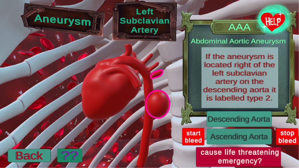
★ My Contribution to Aortic Aneurysm Simulator ★
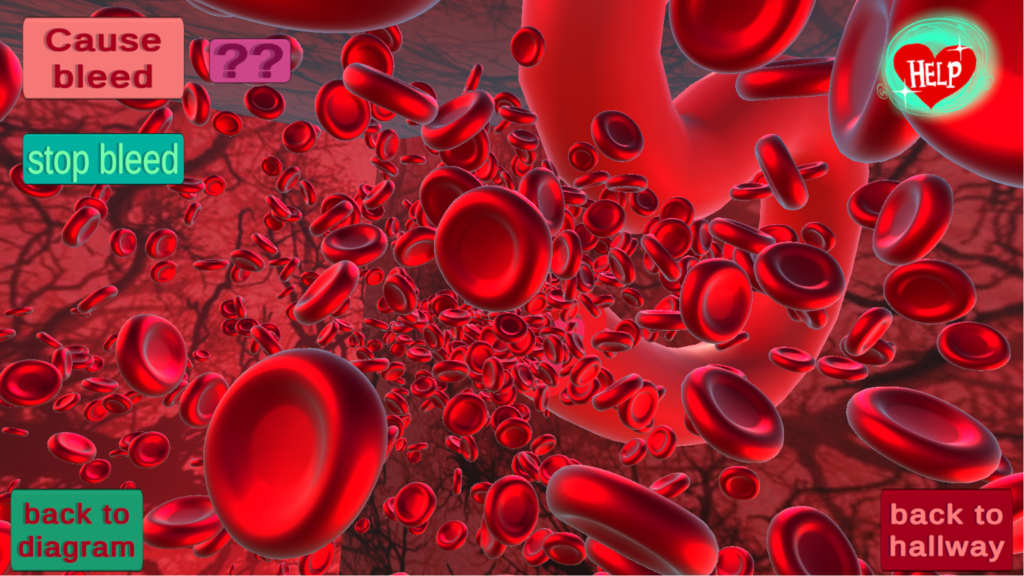
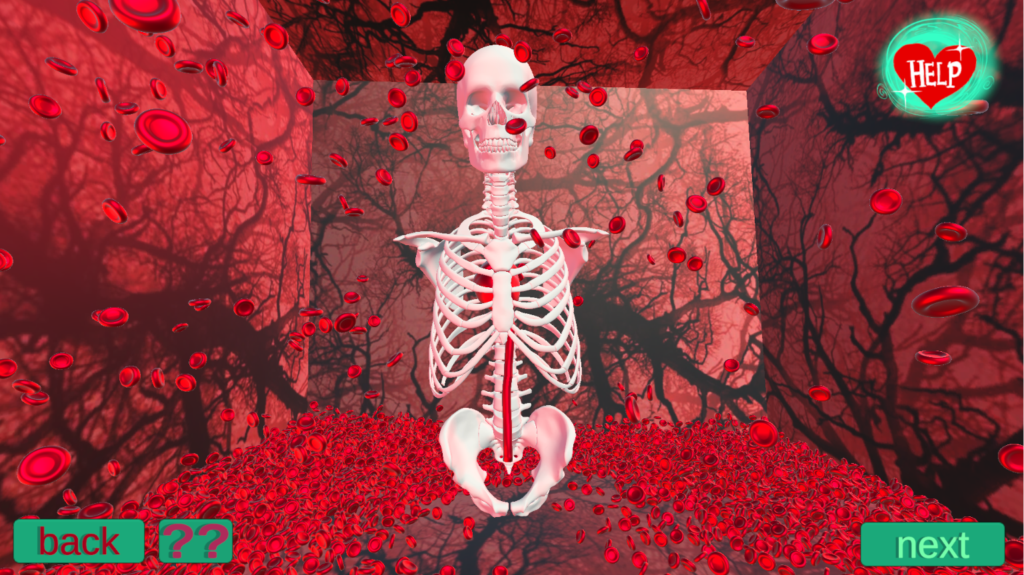
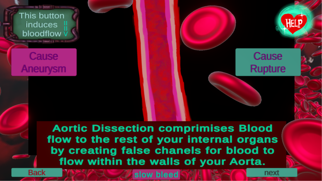
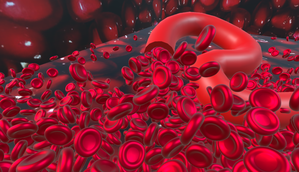
Throughout this project I found creative ways of reusing the same base format for the design of each room to reproduce different user experiences utilizing 2d and 3d animations and graphics. I particularly enjoyed this blood spawner function, creating a script that continuously instantiates red-blood cells to produce a deluge effect. This recreated the imagery of internal bleeding in a visually lighthearted manner. Although this wasn’t a necessary feature for the objectives of this project as an educational application, personally think the BloodSpawner is an engaging and entertaining aspect of the application.
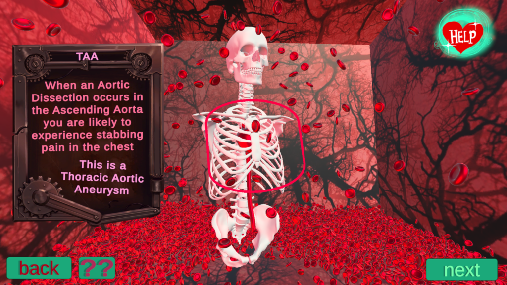
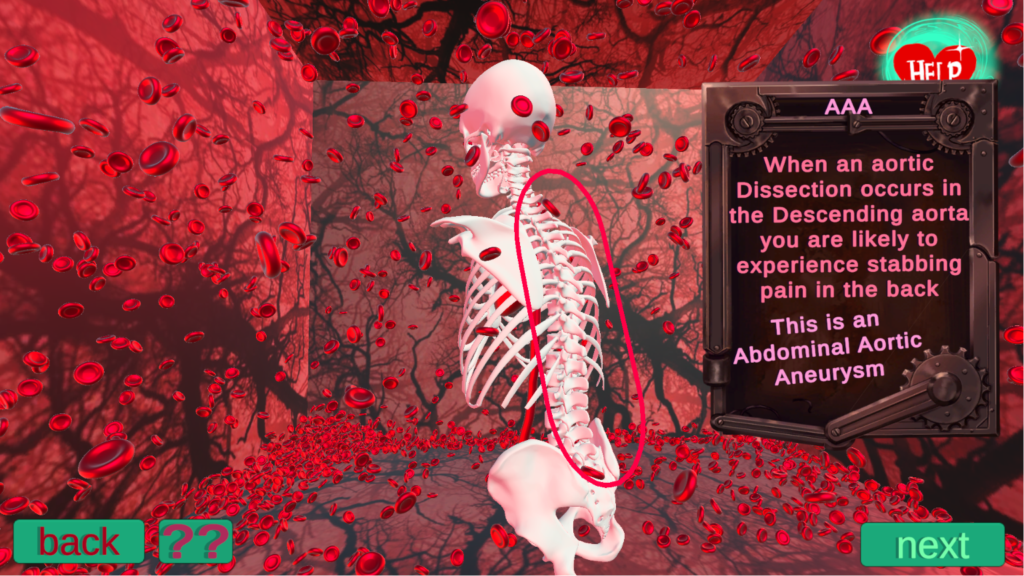
I similarly enhanced the visuals by using interesting graphics and Pomegranate textures for the backgrounds in the playable environments, to create the illusion of human organs in the background. I made use of multiple camera angles and 3d objects to create visually and spatially distinct environments for the educational experience.
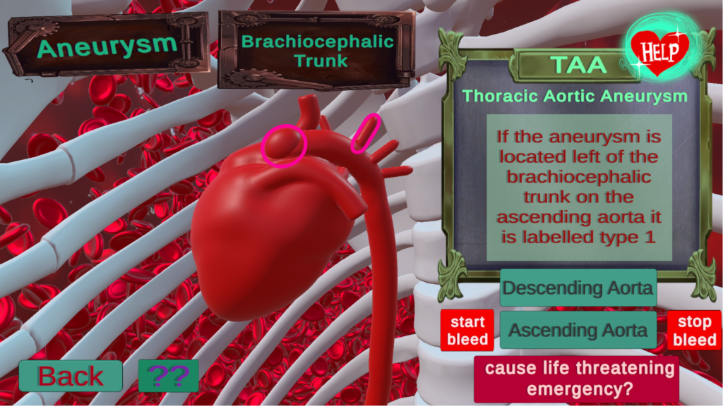
For one of the scenes where the player is to rotate a 3D heart model I placed the camera inside of the ribcage and placed a low opacity red panel over the camera to act as a filter. The intended effect was to create a red tint to create the user experience of being inside the human body.
I put a lot of heart into this project.
Aortic Aneurysm Alert (AAA!)
An Educational Game for the General Public
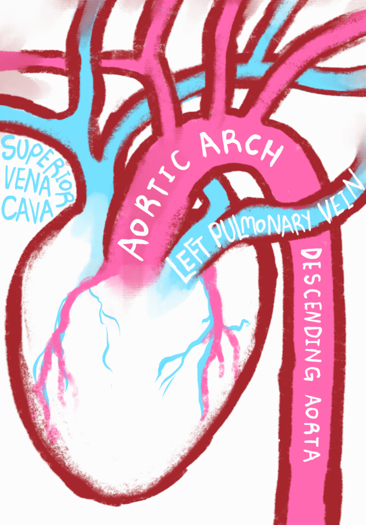
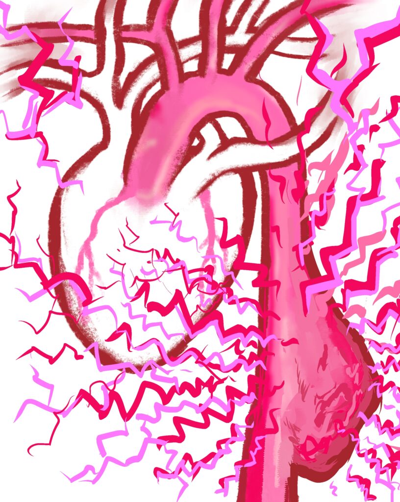
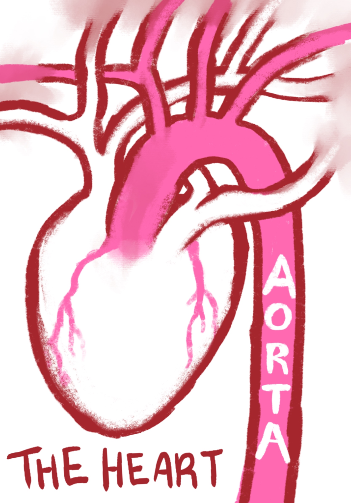
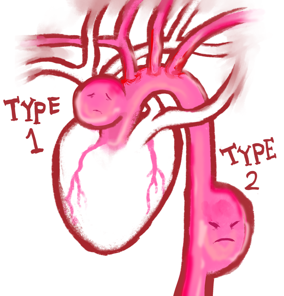
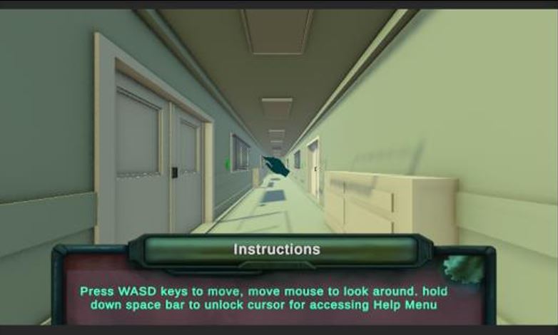
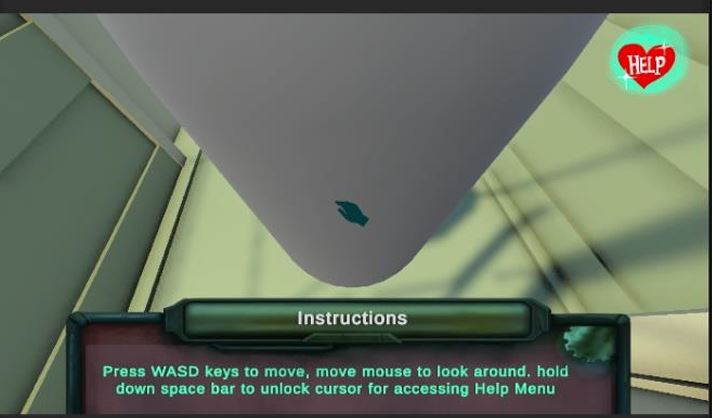
Aortic Aneurysm Alert is a collaborative Unity application, with my amazing and talented course mates Laura Hudson and Precious Karley, designed to make learning about aortic aneurysms more visual and engaging. It allows users to explore a series of themed rooms with 3D anatomical models, animations, videos, interactive activities and a final quiz. My main contributions included modelling and developing the main corridor, creating a physics-based player controller for the player to move around the room, and an interactive ray casting feature where the user can click on the buttons in the corridor and enter each door which is a new scene, and building a terminology scene where users can rotate, inspect and highlight anatomical structures of the heart, as well as learn some information about their heart. The final experience combines anatomy, storytelling and gameplay to communicate medical information in an interesting way to the general public.
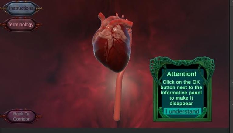
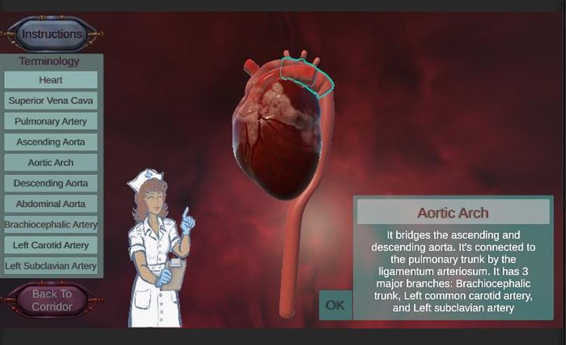
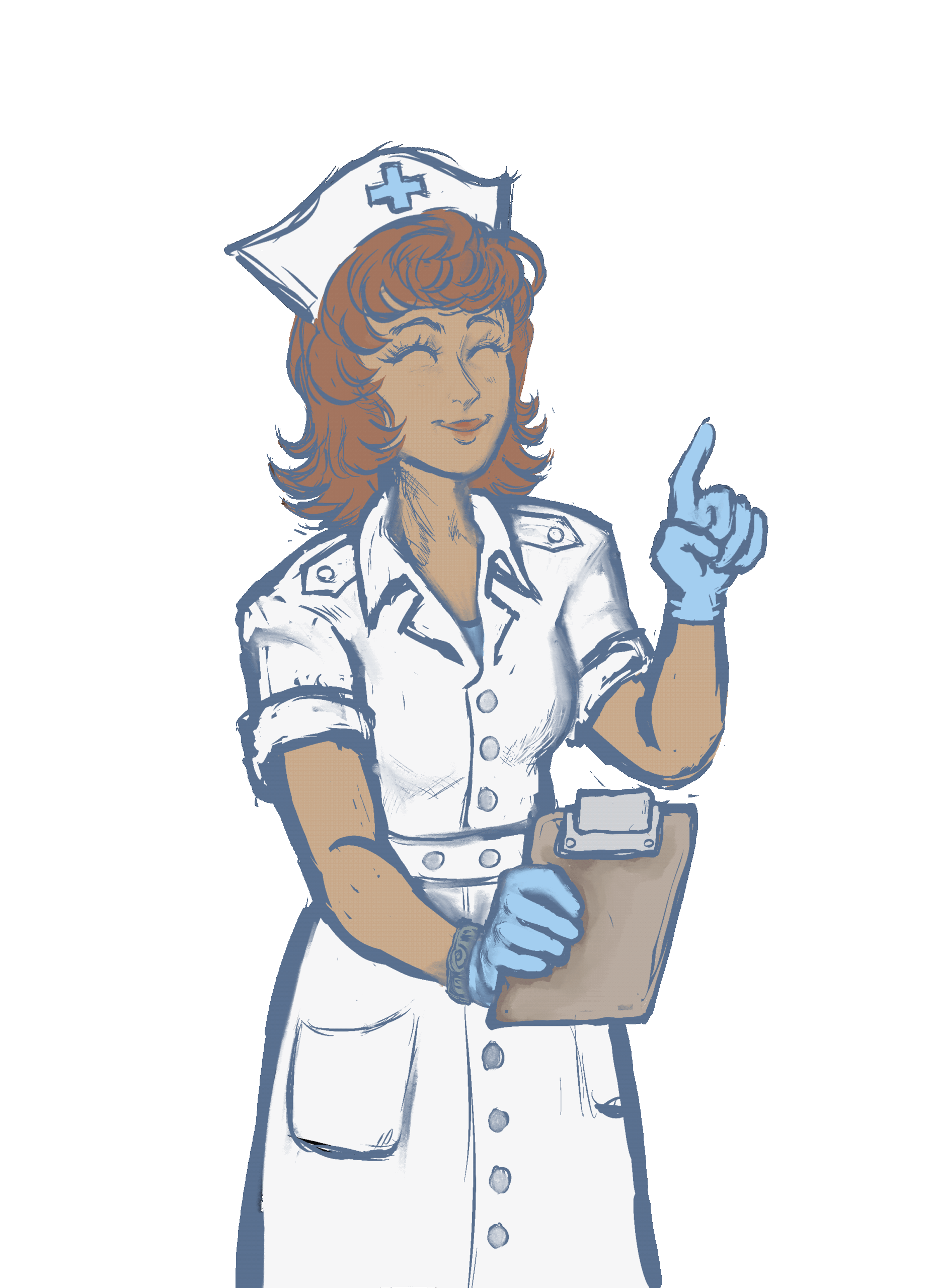
note: the nurse drawing was made by Precious Karley