

Yogitha Chukkala
(She/her)
Hello! I am Dr Yogitha Chukkala. I am from a coastal city called Visakhapatnam, India. I completed my undergrad in dentistry at GITAM Dental College in India in 2022 and practised dentistry until Oct, 2024. I have been passionate about animations and front-end design ever since high school, and thus the decision to do my MSc. in Medical Visualisation and Human Anatomy. I have loved my journey here at GSA and at UofG and will always cherish every step I have taken. A few pieces of work I have created during my time here are displayed below. I do bring that special mix of health education and health care application in every part of my work. Hope you find them intriguing, the way I felt while creating them.
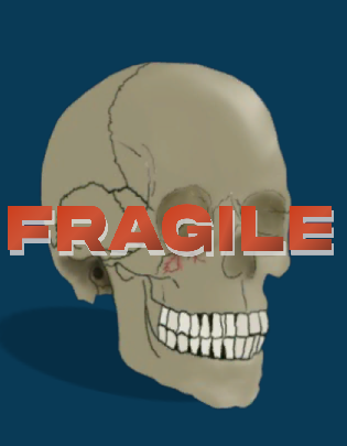
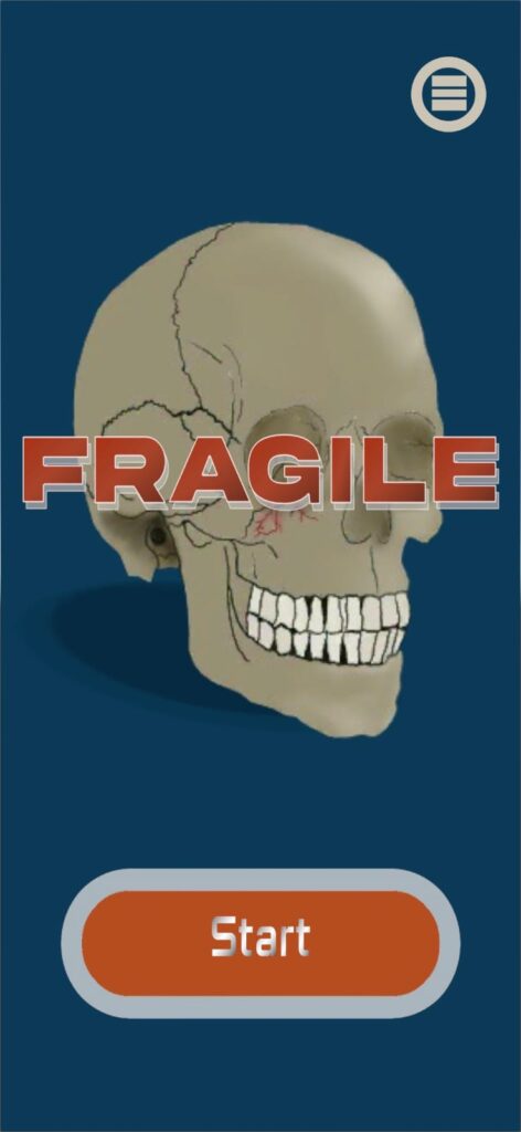
FRAGILE: A Health Education Application with AR Double Vision Viewer
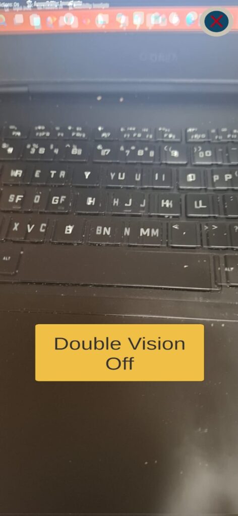
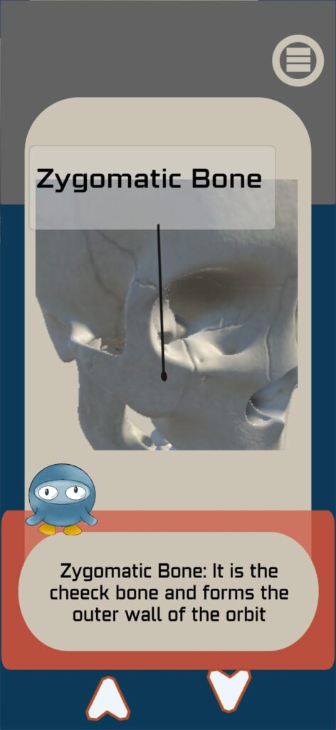
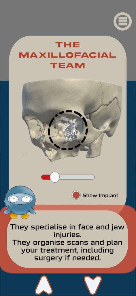
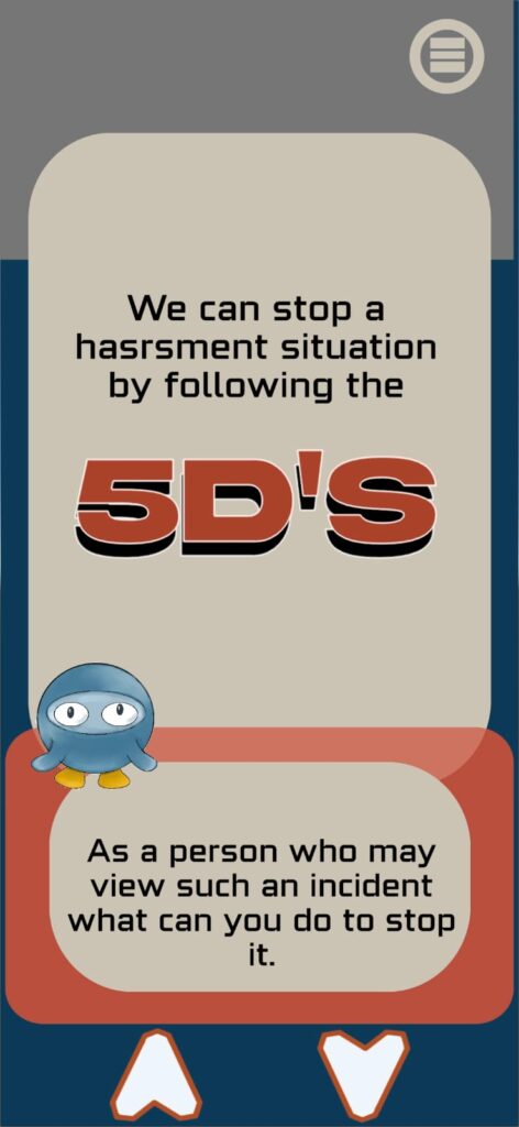
Fragile is a gamified educational mobile application designed to teach teenagers aged 13 to 17 about the physical consequences of facial violence, specifically orbital floor fractures and diplopia. Built in Unity 6 for Android, the application guides participants through three sections: Explore, which delivers interactive clinical content including 3D orbital fracture models and an augmented reality simulation of double vision; Apply, which introduces the 5Ds of Bystander Intervention; and Evaluate, a scored quiz with a tiered badge reward system. Orbis, a custom-designed instructional character, guides participants throughout, making complex medical concepts accessible, engaging, and memorable for a teenage audience. This was made with the supervision of Professor Christine Goodall and Dr Matthieu Poayde.
A video explaining the application: https://drive.google.com/file/d/1i6BVGiHOEH-YhKu4Sw_AvFUSVE7iGpKS/view?usp=drive_link
Volumetric Visualisation
Segmented and registered the given CT and PET data and rendered them into 3D models to showcase the data for patients
What did I do:
- Cleared, filtered, and segmented pre- and post-op CT of fractured hip bone and implantation
- Direct and indirect rendering of the teeth showing its anatomy
- Registered CT, MRI, and PET data to align and render output showing brain tumour, its boundaries, metastasis, and anastomosis
- Segmented lung CT data to show lung cancer on both lobes
Result: Four rendered outputs of four different anatomies and their datasets.
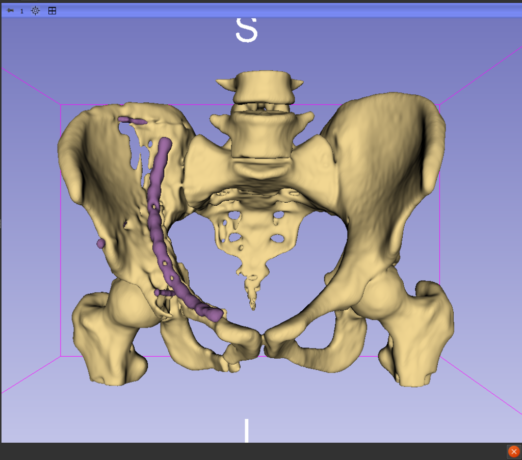
Post op Pelvic bone segmented showing the Implant
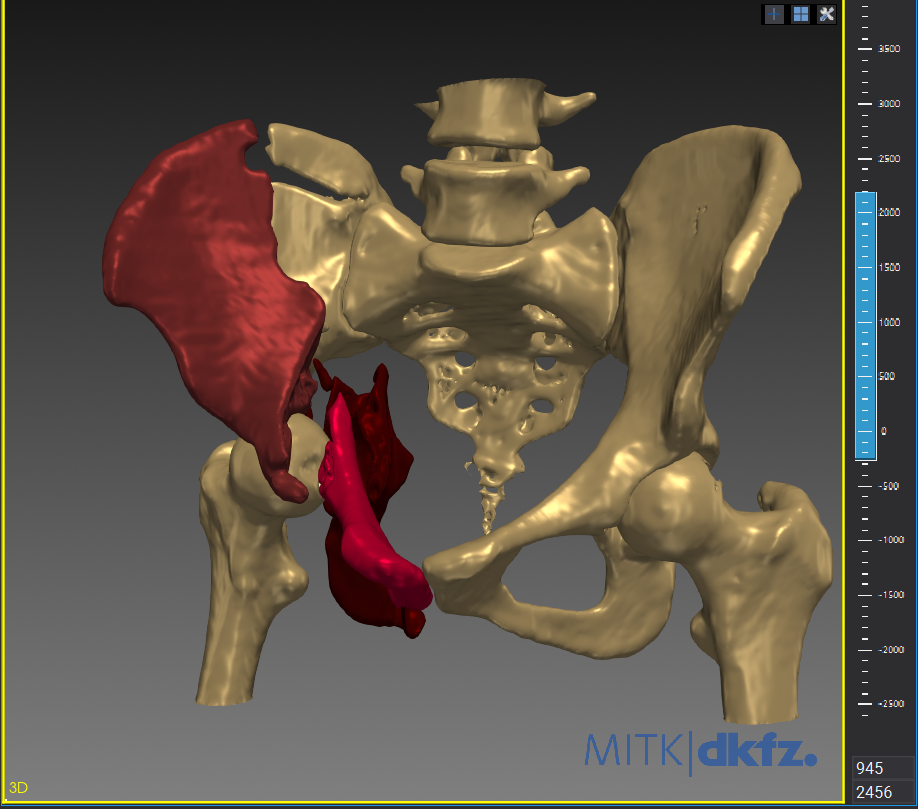
Pre Op Pelvic bone showing the fractures segments
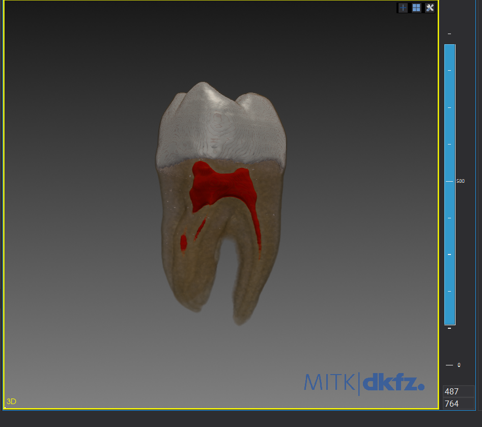
Direct Volume Rendering of Teeth
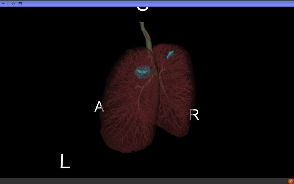
Lungs showing tumor growth on both the upper lobes
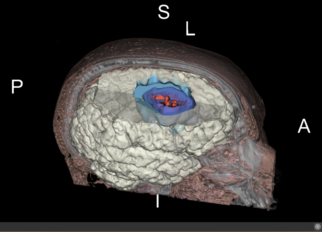
Brain Tumor showing the growth, vasculature, metastasis and borders
Know Your Tooth – Thumb Sucking & Dental Development Teaching Interactive App
It was a collaborative and coordinated effort on an interactive interface for educational purposes, in this case to teach children the consequences of thumb sucking. This was made with the interest of teaching participants about thumb sucking and deformation of the teeth.
Steps that we have done
- Coordinated a cross-functional student team, aligning members on educational objectives and session structure; contributed clinical expertise in paediatric dental development.
- Designed scenes showing the animation of thumb sucking and how teeth get misaligned, controlled using a slider
- Sourced models from Sketchfab, retopologised and corrected accordingly
- Used Unity for animations and C# scripting to control the slider mechanism and buttons
Result: A part of the project showing animations of a child sucking their thumb, how teeth get misaligned, and what a parent should do to educate the child and stop the habit.
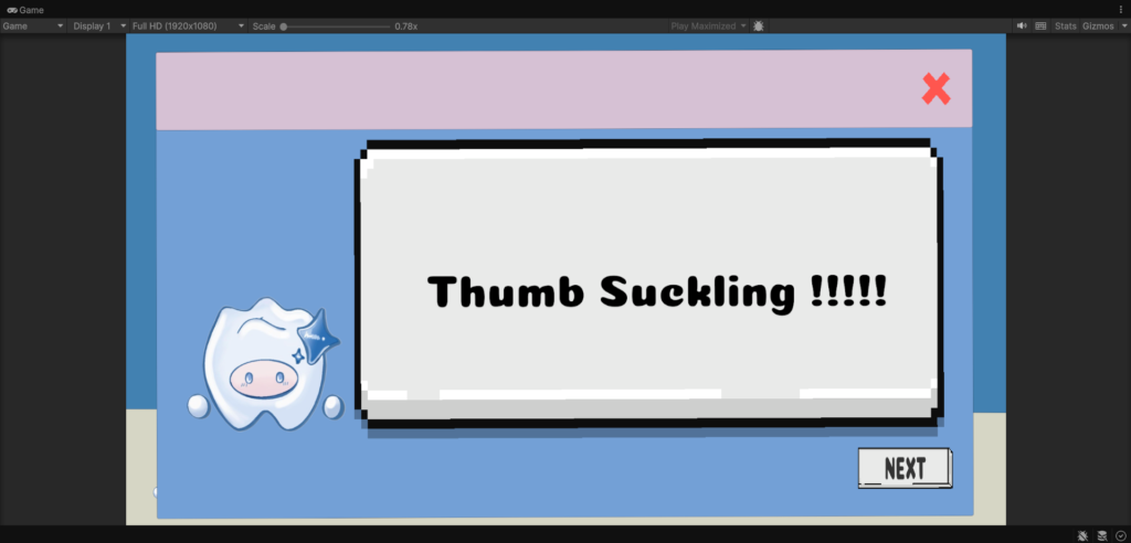
Thumb Sucking Scene that is a part that I have developed
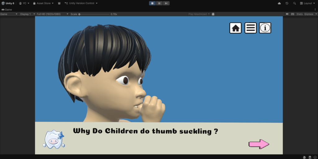
3D model Animation showing thumb sucking
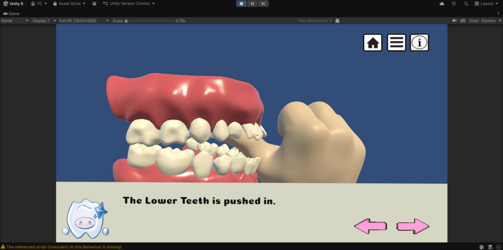
3D model Animation showing thumb sucking and movement of the teeth
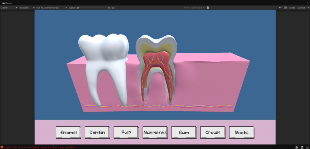
3D model that was taken from SketchFab and edited by me this is used in the Anatomy Scene
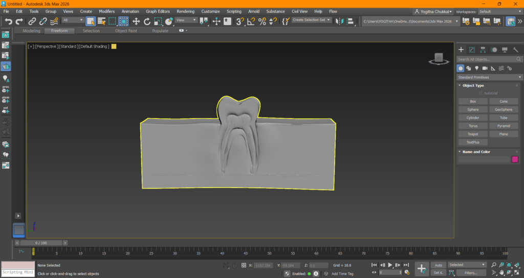
3D model that was taken from SketchFab and edited by me
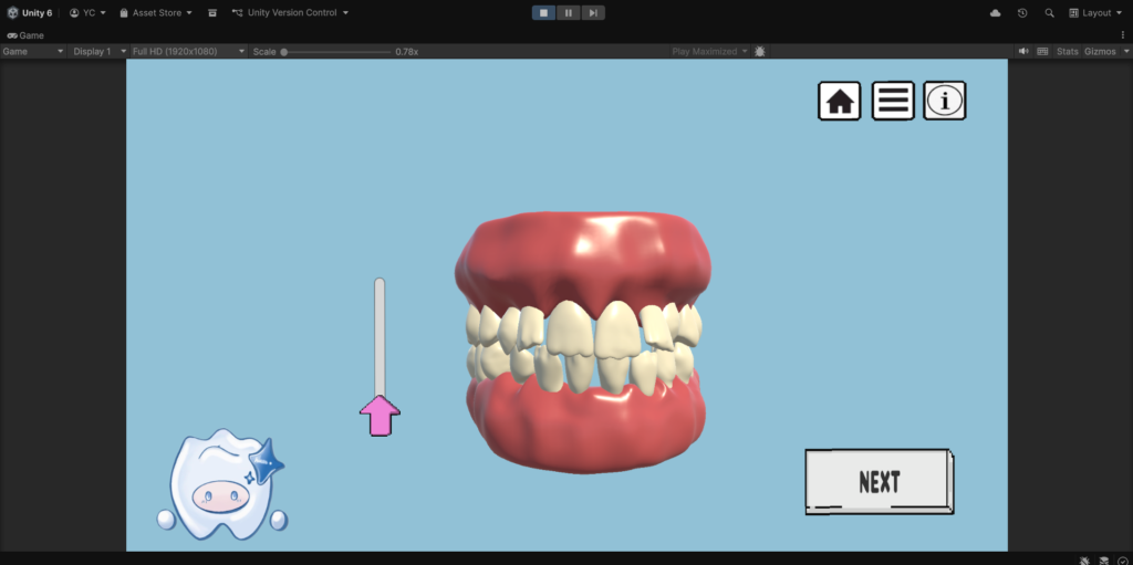
Slider scene that controls the movement of the teeth
Upper Limb Anatomy & Bharatanatyam Mudra Animation (3D Modelling Sumative)
This project was a 3D modelling summative assignment. It is a passion project combining muscle movements and an Indian dance form, where I have created an animation showing movement of the arm and hand bones and muscle contraction while performing certain mudras
Steps I have taken to achive this:
- Retopologised and modelled the complete right upper limb skeleton, including all 27 hand bones, using Adobe 3ds Max and Maxon ZBrush
- Sculpted the Flexor Digitorum Profundus from scratch in ZBrush
- Created alphas and textures for bones and muscle using Adobe Photoshop and Illustrator
- Animated the hand performing Bharatanatyam mudras in 3ds Max, showing joint mechanics, tendon attachments, and anatomical range of motion, edited in Adobe After Effects
The result was an animation video showing the Bharatanatyam mudras, demonstrating precise understanding of joint mechanics, tendon attachments, and anatomical range of motion. I have learnt a lot through this project, not just about animations and 3D modelling but also rendering, video editing on Adobe After Effects and Camera work.
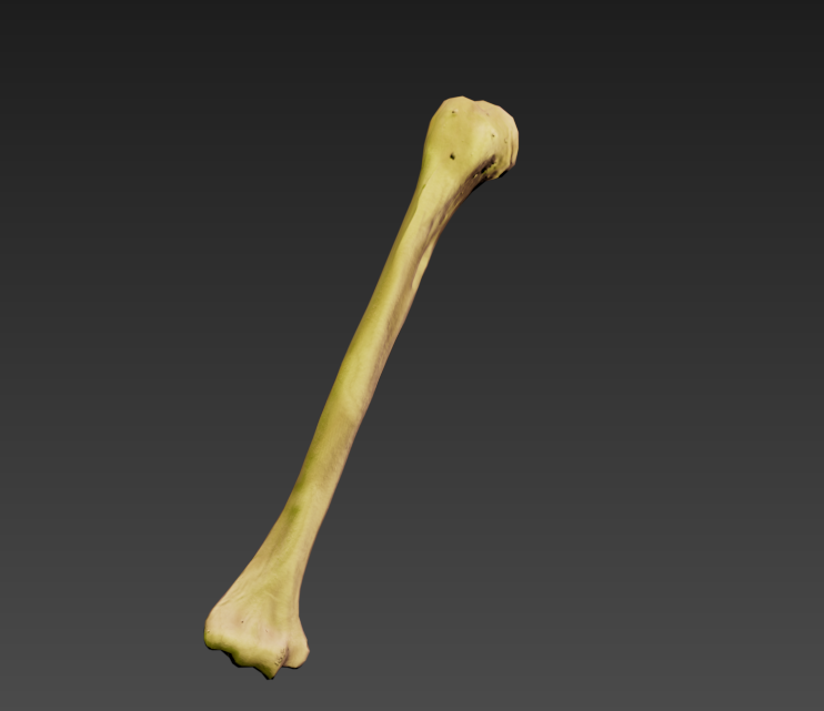
Retoplogised and detailed humerus bone
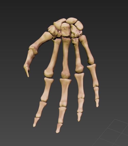
The hand- Retoped and detailed
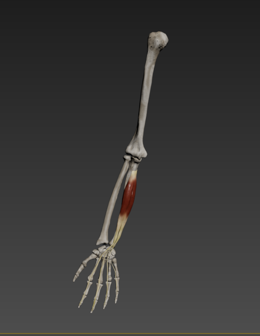
Complete Arm
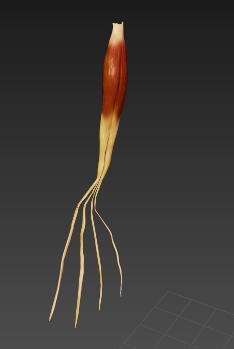
The FDP muscle modelled from scratch and detailed
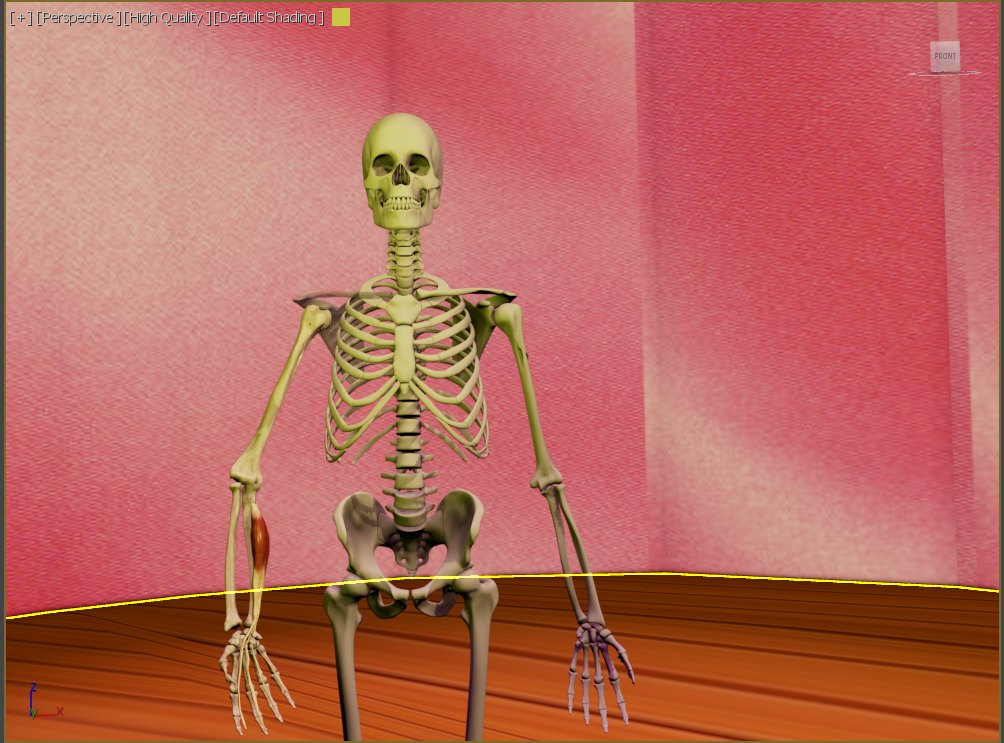
The complete staging
Temporal Bone Learning Interface
My first-ever Unity Project where I have made an interactive interface to teach the parts of the temporal bone.
The process I followed in creating this project was:
- Learned about the temporal bone and its significant landmarks
- Modelled the temporal bone from scratch using Adobe 3ds Max and Maxon ZBrush.
- Created a Unity interface with panels explaining the temporal bone, a rotatable 3D model (C# script), and widgets showing info on 7 important landmarks
- Added a quit panel to exit the game
The overall interface had an introduction and a part which shows the 3D model of the Temporal Bone, where the viewer could view its anatomy and a list of buttons that show information regarding parts of the temporal bone. The project was made to teach the viewer about the temporal bone.
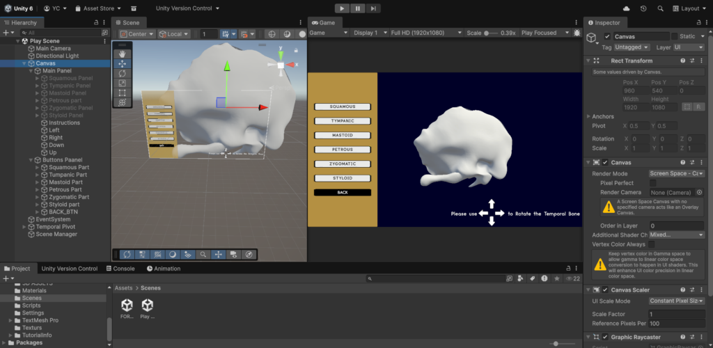
The scene shows work on the Temporal bone Viewer Scene on Unity
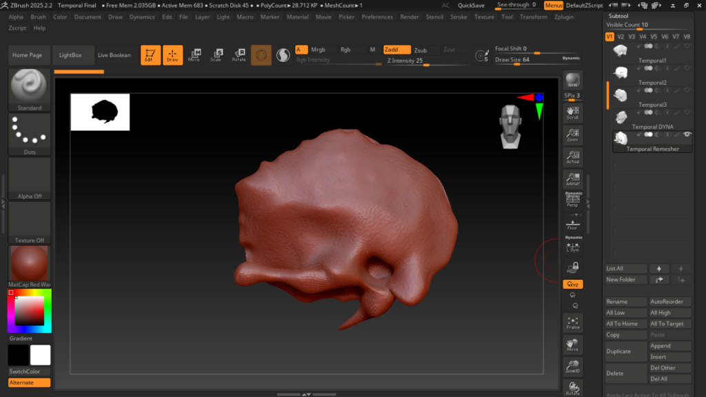
Temporal Bone 3D model on ZBrush while modelling
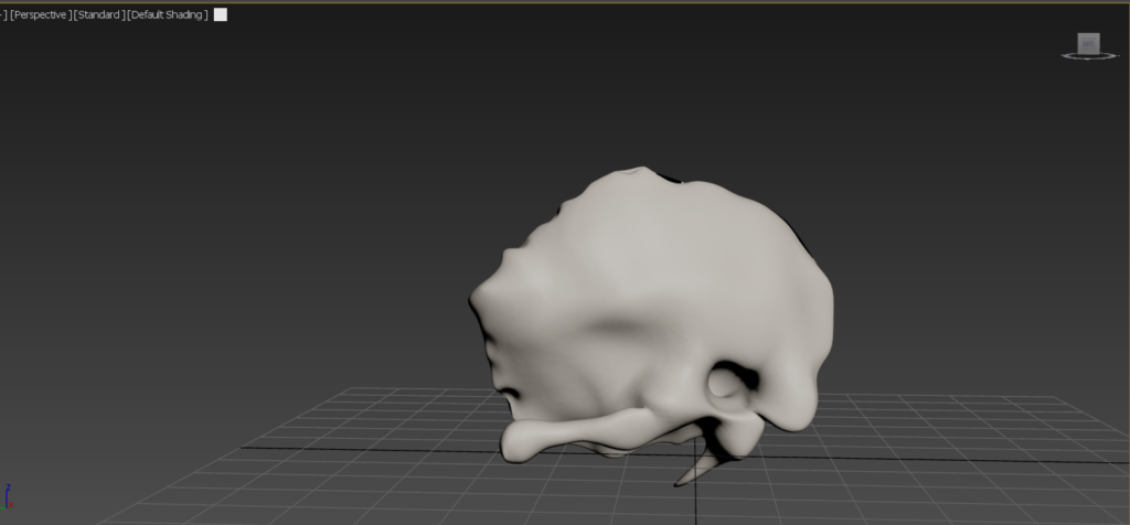
Finished Temporal bone model on 3Ds max
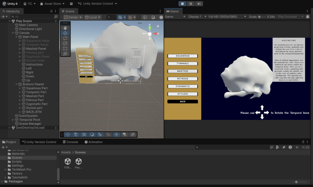
The Temporal bone Viewer Scene showing the panel with information
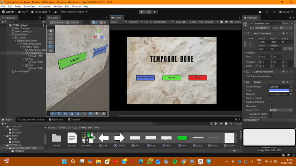
Start Scene