Georgena Buesnel

Hello, my name is Georgena Buesnel. I am an Anatomy graduate (BSc Hons) from the University of Glasgow with a passion for learning about the human body, but also, keen to get involved in tech. This led me to the Medical Visualisation and Human Anatomy degree which perfectly combines the two!
Throughout the course I have gained skills in volumetric visualisation using scan data, 3D modelling, game development, and have deepened my understanding of the human body even further through cadaveric dissection and viewing protected material.
Having studied throughout the pandemic I have seen first hand the need for accessible, 3D resources in the world of medicine and anatomy and look forward to seeing how I can help implement this as I begin my career as a Simulation Engineer.
OP 3D
Children with Duchenne Muscular Dystrophy (DMD) are at risk of developing vertebral fractures due to steroid-induced osteoporosis. Although the concept of long bone fractures seems to be well understood by the general public, the morphology of vertebral fractures and development of osteoporosis is not.
The benefits of 3D models, and interactive applications in medical and anatomical education is well-documented, as well as the use of digital resources for patient education. However, there is a lack of a combination of the three, and importantly, there appears to be no 3D resources regarding vertebral fractures.
Therefore, I developed OP 3D, an interactive, web-based application aimed at the parents/carers of children with DMD, with the aim of increasing awareness of key aspects of the conditions.
Model vertebrae were created through segmentation of online example datasets from 3D slicer and bone density models were created from scratch using inspiration images in Z-Brush. The models were refined in 3DS Max/Z-Brush to simplify, texture, and animate the models. The application was developed in Unity to collate the UI elements, 2D, and 3D animations and develop interactions between the user and 3D models.
OP3D was uploaded to itch.io for user testing and was advertised on DMD patient organisation groups. A pre- and post-application awareness questionnaire was developed to assess how use of the application affected participants awareness of key aspects of the conditions. In addition, the standardised System Usability Scale questionnaire was utilised to determine the usability of the application as a whole.
Thank you to Dr Matthieu Poyade, Dr Jarod Wong & Ibrahim Buksh for all their help with this project.
Project Links


Volumetric Visualisation
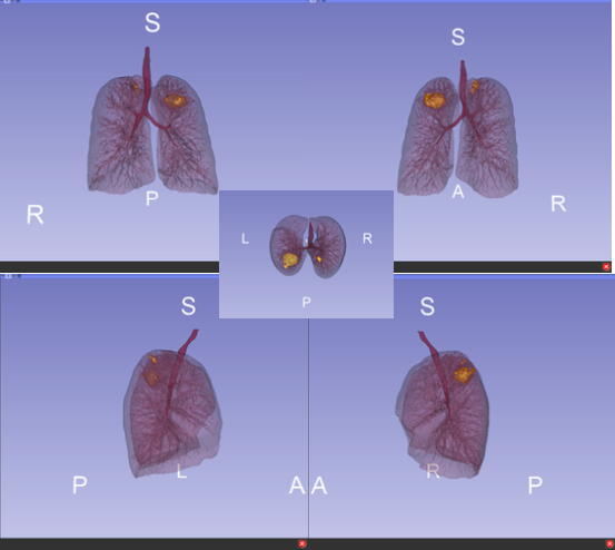
Volumetric Visualisation is a module of the Med Vis course and teaches students about the creation of 3D renders from 2D, traditional scan data such as CT and MRI scans.
I gained skills in both indirect and direct volume rendering as well as data registration (the alignment of data) and produced a high quality assignment utilising these skills. This assignment provided me with a deeper understanding as to what technique to choose when faced with a problem, a skill which I adopted during the development of my MSc project when creating vertebrae models.


Interactive Visualisation
Limbic Learner was an app developed by myself and my peer, Ryan, to aid higher education students learning about the limbic system. Both Ryan and I found this a difficult topic in our undergraduate degrees and decided it would be beneficial for some type of 3D resource to be developed catering to this need.
Whilst I used 3D slicer to develop a brain model, Ryan created the limbic system in 3DS Max, both of which were imported into the app. We developed a teaching scene to educate students about the different parts of the limbic system and both a quiz and drag and drop test to assess their knowledge in a fun, interactive and engaging way.


Teach Scene
3D Modelling
The 3D modelling module allowed us to gain experience using 3DS Max to create models from scratch. The final assignment included performing both automatic and manual retopology on a vertebra model as well as modelling the upper arm.
Having not came from an artistic background I found this module the most difficult but also very interesting. It was great to see how myself and my peers created such different models from the same source image! It was also extremely helpful to combine this skill with my existing knowledge of human anatomy which I believe helped me understand the mechanisms of the upper arm better.
